My 00:02:23 of Fame

by Jennifer Frazer on April 13, 2011

Over at the podcast This Week in Virology (TWiV) at virology blog (itself long a feature of the Artful Ameoba blogroll), this blog was kindly chosen as his Pick of the Week by Alan Dove. Since perhaps not all of you are interested in listening to an hour and  a half of virology, the bit about me starts around at 1:26:10 and runs to about 1:28:33

For a person who started out doing this blog (and still does it) sitting on her futon in her jammies, I can’t tell you what a thrill it is to hear your blog discussed on the radio! Thanks for the pick and the kind words, guys!

And in case any of the TWiV guys are reading, once I figure out what the chromatoidal bar of the cyst of Entamoeba histolytica is, I will definitely consider doing a post about it. : )

{ 5 comments }

The games in this video could be used to teach about biology, according to the folks at New Scientist. Or about behaving as a merciless puppetmaster/God to a bunch of innocent protists. Your choice.

I’m torn here. On the one hand, this is really cool. On the other, it somehow feels horribly wrong. I mean I’m sure it’s not much more wrong than what I do to baker’s yeast every time I bake bread. But . . . I don’t toy with the yeast before I incinerate them. Yeesh.

Anywho, for those who are unfamiliar, paramecia are classic organisms used to teach high school and college students about microbes. But they’re not simply boring microbial lab rats. They’re fascinatingly formed, filled, and operated little creatures. They are often described as “slipper-shaped”, or, as van Leeuwenhoek would have it, “slipper animalcules”. In this video, watch for the little cilia beating all over the cell’s pellicle, or membrane+submembrane shell.

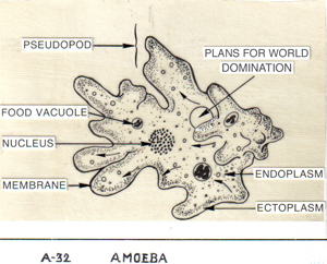
So let’s have a tribute to the humble Paramecium today — we’ll take a closer look at a microbe you might think you already know. Think again. Take a look at the basic paramecium formula here (I can’t post it because I don’t have copyright). I’ll refer to the various parts of this diagram as we go along.

Paramecia are ciliates, protists coated in little beating hairs called cilia for at least part of their life cycle. In photographs where only their cilia are stained or photographed with scanning electron microscopy, paramecia look like ovoid shag rugs (or see here or here). These cilia can beat in forward or reverse with extreme precision — much more precise than large flagellae or lumbering pseudopods. When beating in foward or reverse, the paramecium moves in a spiral motion around an invisible axis — and they can throw it into reverse in a ciliumbeat to avoid obstacles or “negative stimuli” (cough).

Inside the cell is a contractile vacuole used to bail out water against the perpetual osmotic gradient (the inside of the cell has a higher solute concentration than its watery surroundings, leading water to constantly seep through the membrane by osmosis, like a leaky ship), various vacuoles (spherical storage vessels) for digesting food and excreting waste, and at least one micronucleus and macronucleus.

The micronucleus, of which there may be as few as one or as many as 80, is diploid, or contains two copies of all the DNA, just like all of your cells. But the macronucleus may contain 50-500 times as much DNA as the micronucleus (the Paramecium aurelia macronucleus is 860-ploid, according to one of my biology texts). The micronucleus is all about passing DNA to mating partners for sexual reproduction (aka swapping genes), while the macronucleus is in the business of pumping out messenger RNA and getting it translated into proteins.If you remove the micronucleus, the cell can divide asexually another 350 or so times before dying of no sex (yes, in ciliates, this can apparently happen). But if you remove the macronucleus, the cell immediately dies.

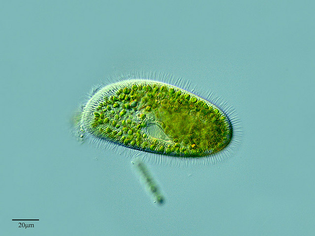
On the surface of the cell is a funnel-shaped oral groove that guides food particles toward a pocket where food vacuoles are created. Enzymes get pumped inside to digest whatever hapless bacteria, yeast or “other” finds its way inside. Many paramecia may contain symbiotic bacteria living within them or even within their macronucleus, perhaps providing vitamins or other growth factors that would otherwise be hard to get. One paramecium even has photosynthetic symbionts. Paramecium bursaria, which has ingested and partnered with the green alga Zoochlorella.

These are *not* chloroplasts, or rather, they are not homologous to (share a common ancestry with) the chloroplasts in plants. Plant chloroplasts are ingested cyanobacteria. Zoochlorella are chloroplasts in the sense that they are internal photosynthetic symbionts, but they were free-living eukaryotic (nucleated — not bacterial) algae before ingestion. But here’s the mind-twister: the chloroplasts within the Zoochlorella are homologous to plant chloroplasts, because plants evolved from green algae.
This is the only Paramecium known to do this. To what extent the Zoochlorella could pop out of that paramecium and get along on its own or is degenerate and helpless I do not know.

Paramecium bursaria, a rare symbiotic photosynthetic paramecium. Love the gorgeous detail. Creative Commons PROYECTO AGUA**/**WATER PROJECT

Some large ciliates may hold the land-speed record for all protists. They can cruise at a blinding 2 millimeters per second, which, assuming a 250-micrometer paramecium and a 5.5-foot human [calculator tapping noises], is the equivalent our 5.5-foot human swimming  a blazing 30 miles per hour*. And they are backstroking at these speeds through a medium that has the viscosity (to a proist) of molasses.(See the middle of Psi Wavefunction’s post here, or for the physics-buff bionerds out there, the original 1977 paper “Life at [a] Low Reynolds Number” here).

Paramecia are also armored with a built-in defense system. Their pellicle is laced with dart-tipped harpoon-esque “trichocysts” (a specific type of a more general organelle called an “extrusome“, which is clearly another case of convergence (independent invention of the same structure) with jellyfish nematocsyts), which deploy explosively within milliseconds if the paramecium feels threatened, is feeling peckish, or perhaps needs a convenient anchor while it tucks in to dinner. See how they look retracted here, and how they look once fully-expelled here, which this site describes as the “disheveled porcupine” look. The bottom photo is of undeployed trichocysts in the pellicle**.

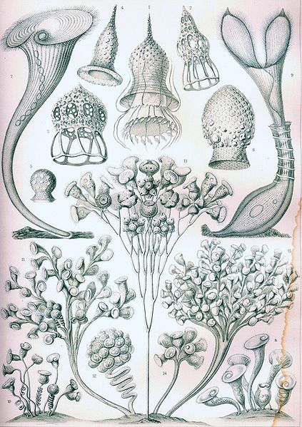
Known diversity in ciliates, ca. 1904, by the incomparable Haeckel

Paramecia are, as mentioned, ciliates. There are something like 9,000 species in fresh and salt water, and their complexity and diversity of form is astounding. Some of the most complex species come close to replicating the digestive systems, muscles, exoskeletal systems and even vertebrae so characteristic of multicellular life within a single cell. According to one of my biology texts, ciliates have produced the greatest specialization of subcellular organelles of any protist. Rest assured you will read more about the creatures in this group at this blog.

Ciliates fit into the larger taxon (group) called alveolates, a term you may recognize from high school biology as the name for the little sacs at the ends of the passages of your lungs. The structures are similar; the Latin just means “little cavities”, and in ciliates like Paramecium they are little membranous sacs found just below the cell surface engulfing the roots of cilia. Though the ciliates like Paramecium and the dinoflagellates and parasitic apicomplexans may seem worlds apart, they all share this feature and several others*** that reveal their common ancestry. See how the ciliates fit into the alveolates, and how the alvaeolates fit into the eukaryotes here and here.

Before I leave you today (or is it the other way around?), enjoy this video of some more active paramecia tooling around with a bunch of nerdy Euglena (the little guys). Because before you allow your little microbial minions to be devoured by Pac-Mecium or taunt them with a chemical blast, remember: were they your size (or you theirs), you’d never survive the hairpin extrusome assault mechanism, and they’d be gone before you knew what hit you. : )

________________________________________________
*or 48 km/hr for you metric users. According to the intertubes, the fastest human swimmers can go is around 5 miles/hr over very short distances. Of course, their bodies are not covered in highly-efficient propulsive cilia (and more’s the pity).

**With all this weaponry, forget Pac-mecium. Helloooooo Ultimate Paramecium Fighting League.

***including, for the bionerds out there, tubular mitochondrial christae, “closed mitosis” in which the micronuclear membrane doesn’t dissolve during mitosis and the mitotic spindle forms inside it, and similar “extrusive organs” like trichocysts

{ 7 comments }

Graptolites Have Tentacles Too

by Jennifer Frazer on April 1, 2011

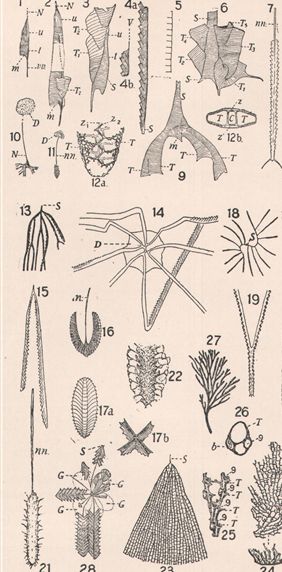
A graptolite sampler, from an ancient Encyclopedia Britannica

Occasionally, life looks like it isn’t. In the eastern forests of North America and in a thin strip along the Pacific Northwest (but sadly not in Colorado), hidden in plain sight on tree trunks you can find the gracefully named elven script lichen, Graphis scripta. With a little imagination, the lichen looks like secret writing, not like an eruptive fungal-algal symbiont that specializes in cohabiting in tree bark.

In the 18th century, Linnaeus, the father of taxonomy, faced a similar biological dilemma. He found patterns in rock he suspected were chemical or geological formations that looked as if they had been alive, but actually had not. He called them “graptolites” (= rock writing), and gave the name to a variety of things may or may not have been alive. Over time, however, the term was co-opted by paleontologists for a group of strange fossils that very much had been alive. The only problem was, no one knew what the living parts of these things actually looked like — or how they might be related to anything alive today.

At left, you can see a variety of these creatures’s tube-like houses, called coenecia (se-nee-see-a), which are found abundantly through marine rocks from the Ordovician, Silurian, and part of the Devonian. Some were branched and tree like (dendritic, see13, 18, and 27, left) and probably bottom-dwellers, and others took on a variety of other bizarre forms that scientists interpret as the products of a planktonic form.

Many tended to have characteristic “hacksaw” shapes (see 4a, 7, 15, and 19, left) either in tuning forks, or coiled up in spirals like watch springs, as if something had poked out of the teeth lining these tubes. But no one knew what. The few cases where some actual ex-animal had fossilized were apparently more like ex-animal smudges than ex-animal fossils.

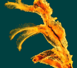
The pterobranch Rhabdopleura, in a lovely study in blue and gold. Note the tentaculate feeding/breathing prongs, aka lophophores.

Meanwhile, in another part of the science universe, scientists were describing and identifying members of a group called pterobranchs (= winged or feathered gills). Stretching little more than a centimeter long and living in proteinaceous* banded tubes cooperatively secreted by their shield-shaped probosci, they humbly go about their business stretching their ciliated tentaculate arms (which may remind you of bryozoans’lophophores, which they merely resemble convergently because that’s what they probably are (see comments)) into the water currents to catch prey and exchange gases. Inside their proboscis is a true lined body cavity, or coelom (seel-um). They sometimes live on their own, but usually grow in colonies connected by stems, or stolons**, in that colony of fused tubes called the coenecium. Some species have a pair of gill slits, just like fish (For a nice look at the general structure, see here, here, or, yes, the plush version here).

And this may very well reflect pterobranchs’ position in the shrub of life. Pterobranchs, it turns out, are hemichordates, in the same group as the acorn worms (enteropneusts) I described here last year. They are animals that evolved from animals just on the verge of becoming chordates, or nerve-corded animals like ourselves. They have a tripartite body plan of proboscis, collar (whence the tentaculate arms spring), and trunk. Like echinoderms (seastars, etc.) and chordates (us, etc.) they have a complete digestive tract whose mouth forms from the second indentation in the hollow ball of cells formed after a fertilized egg starts dividing (= deuterostome). Like echinoderms (but unlike chordates), they have no body segmentation and a special kind of larva called a dipleurula (chordates have no larvae). Signficantly, a hollow neural tube grows in some species early in development.

Most suggestively, pterobranchs and the fossil graptolites seemed suspicously similar, although little more than two dozen species of pterobranch live today, and for millions of years in the Paleozoic (from the Cambrian to the middle of the Devonian), graptolites were the dominant zooplankton in the world’s oceans. Tantalizingly, the microstructure of graptolite fossil tubes is very similar to the microstructure of pterobranchs, a detail discovered when electron microscopes first peered inside tubes of both animals and ex-animals in the 1970s. But no preserved fossil animal could confirm this.

Looks like a graptolite. Quacks like a graptolite. Could be a graptolite. Wouldn’t it be great if we had some soft tissues preserved in a graptolite fossil! Well, now we do.

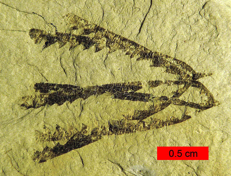
Galeaplumosus, which was probably a two-armed model. The right arm is broken off, but two tentacles are still visible on it. "You don't look a day over 500 million years. You and Rhabdopleura could be sisters!" From Hou et al., Current Biology. Click for link.

In a March paper in Current Biology, scientists report the discovery of a tentaculate graptolite 525 million years old from the lower (early) Cambrian.

Finally, in all its glory, an animal poking out of a conical graptolite tube. And what an animal!For pterobranchs, they are, at shy of two inches (four centimeters), Yao-Ming-class. Which is fitting, because the fossil was found in China and dubbed Galeaplumosus abilus, from galea (helmet) and plumosus (feathered), and ab (away from) and nubilus (cloudy). Yunnan, where the fossil was found, means “south of the clouds”.

The fossil provides the clincher on graptolites’ true identities: a banded (probably secreted) cooperatively-made tube with contractile stalk and tentaculate feeding arms projecting from the opening is the M.O. of extant pterobranchs.

Looking carefully at the fossil, scientists were even able to discern possible cilia (silly-uh — little hairs that beat back and forth to draw in particles of food) on one tentacle, and a possible contractile stalk inside the shell. What scientists have, apparently, is the earliest, largest hemichordate animal (zooid) ever found, alive or dead, and it seems to show that their way of feeding and building a house have changed virtually not-at-all in 525 million years. Take that, sharks***.

The authors of the paper hypothesized that the rarity of specimens like this is probably a result of most graptolites’ planktonic lifestyle: on the long trip to the big sleep, most graptolites/pterobranchs probably decayed before they hit bottom, while the shell has proved decay-resistant in modern tests. That this animal was preserved, they suggest, means it was likely a bottom-dweller.

And that would fit with what we know about graptolite natural history. Scientists long suspected that the first graptolites, which tended to be tree-shaped (dendritic) and evolved in the Cambrian, were likely sessile bottom dwellers. Only later, in the next era, the Ordovician, did a floating planktonic form also emerge, the Earth’s first large zooplankton — and by far the dominant plankton of the early Paleozoic oceans. With their collaborative approach to constructing a floating colony, they were a bit like floating bee hives or wasp nests, if the wasps were all attached at the abodomen by stems, secreted their own cells (instead of building them from chewed up wood or mud), and never left the nest. Like the vast floating chains of colonial salps in today’s oceans (though much smaller), they must have been strange indeed.

The graptolite Pendeograptus fruticosus from the Lower Ordovician (477-474 mya) near Bendigo, Victoria, Australia. This style is referred to as the "tuning fork".

These planktonic co-ops evolved so abundantly and so quickly that they are commonly used as “index fossils” by the geology and petroleum geology sets to date rocks relative to each other with fine detail. In their heyday, thousands of species filled the oceans, common, widespread, quickly evolving and easily identifiable: a rock dater’s dream (errr . . . yeah. Ammonites have also been used this way.) In the Silurian, for instance, 40 different graptolite zones have been described, with an average duration of .7 million years — incredibly fine detail for geologic time, where dating anything to within a few tens of millions of years is usually considered spectacular.

Sadly, the planktonic graptolites went extinct in the middle Devonian, about 380-400 million years ago. Thus, the first (Galeaplumosus et al.) and last forms we find in the fossil record (from the mid-Cretaceous, near the end of the age of non-avian dinosaurs) are bottom-dwelling dendritic forms — as are the handful of species alive today, the humble survivors of a formerly world-dominating group****. But let us take the sunnier view. This post could have been titled “Graptolites Are Not Extinct!”.

_______________________________________________________

To learn more about graptolites and pterobranchs, see here, and a nice page here by the British and Irish Graptolite Group (BIG-G : ) ).

*made of a collagen-esque material, a family of animal proteins that help keep your muscles attached to your bones and your skin perky.

**A term for horizontal connections between organisms. Stolon, incidentally, is also the term botanists use for stems (*not* roots) that run along or jut below the ground from plant to plant (aka runners). If you’ve grown strawberries you have experienced this phenomenon.

***420-million year-old posers

**** much like brachiopods

ResearchBlogging.org
Hou XG, Aldridge RJ, Siveter DJ, Siveter DJ, Williams M, Zalasiewicz J, & Ma XY (2011). An Early Cambrian Hemichordate Zooid. Current biology : CB PMID: 21439828

{ 7 comments }

To celebrate Friday, here’s the best video I’ve ever seen illustrating cellular slime molds, the borg-like creatures that start out as individual amoebae in the soil but then aggregate into a slug that roves around a bit before rearing up into a sporangium, or spore capsule. This particular species seems to be the cellular slime mole lab rat, Dictyostelium discoideum (dik’-tee-o-steel’-ee-um dis-koid’-ee-um). Notice how (apparently) easy it is to find these guys in the soil!

Video is, unfortunately, in German. If you don’t speak German, consider making up your own (PG) translations to key scenes and sharing them with us in the comments! : )

In the beginning you see the free-living amoebae (I think) happily wandering about on their own with some fungal filaments (called hyphae, high’-fee) growing at the top of the screen. Then the ameobae start aggregating — crowding after each other like sports fans filling a stadium. The species uses a famous signaling molecule called cyclic AMP (cAMP) to coordinate their union, and it passes through the swarms in pulsing, spiraling waves noticeable at about 1:35. If I’m using my extremely poor knowledge of German correctly, the narrator is nothing that hundreds of thousands of amoebae join together in the process. They do not fuse membranes; they retain their cellular identity.

Notice that some amoebae get left behind or lost in the process. At 2:47 you can actually see some break out of line and go back to being  little amoebae at the very tail end. After the spiraling and pulsating business is done, the mass stretches into a slug and crawls off. At some point between forming the slug (also called a grex) and making the sporangium (the house where spores are made), the amoebae get it on and mix some genes.

When the slug decides conditions are perfect, it stops, puddles up, and then stretches skyward. The lucky amoebae who will become spores riding up the stalk like an elevator. Those stalk cells get the rotten end of the deal — they must sacrifice themselves to ensure their comrades can reproduce. This little detail has led scientists to study these organisms in order to better understand altruism and cheating in nature. What they’ve found is that, as ever, things are not always as they seem. Some would-be stalk cells indeed give their lives, but others buck the system by cheating. Yet if everyone did, the system would break down entirely. There are, as you may imagine, some very interesting dynamics and mathematics governing this system.

Finally, a roving madsnail goes on a rampage wantonly destroying the beautiful slime mold gardens. Stupid animals.

Incidentally, D. discoideum is the species I wrote about in January in which some strains were recently discovered to practice agriculture, or something close to it, by taking bacteria of their preferred noshing type with them in their spores so they have a guaranteed food source when they land. And still more recently, scientists published an article in Science (see here and here) they may even have tissues — and use two signaling or regulatory proteins related to the ones animals use to organize their embryos during development. This seems to mean the common ancestor of slime molds and animals (whatever *that* might have looked like) was using ancient versions of these proteins to arrange itself, and its descendants — both slime molds, and you — inherited these same proteins and are still using them to organize their bodies, in their different ways.

Cellular slime molds represent one of life’s many experiments in multicellularity. You are the product of another. So are plants. And so are fungi, and brown and red algae and some blue-green algae — and there are many more. Other experiments seem to have been abortive; recently this article revealed that blue-green bacteria (aka cyanobacteria) dabbled in multicellularity many times. Remember: evolution isn’t a goal-directed endeavor, although in certain etremely successful groups (vertebrates, beetles) it may seem that way.

To see a different cellular slime mold species that makes violet sporangia on slime mold candelabra, see here. Spectacular.

Finally, I’d like to note I have a new favorite German word : Schelim. As in “schleim mold”. : )

HT to this post at Small Things Considered for the discovery of this wonderful and educational German film.

{ 3 comments }

Creative Commons Jeans_photos, photo by Fred Hort. Click for link to license.

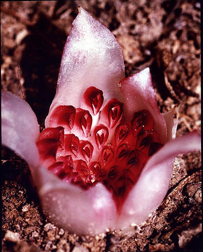
In 1928, a farmer in Western Australia named Jack Trott was plowing a field newly carved from the Outback by fire. An unusual crack in the soil caught his attention. In it was something extraordinary — a sweet-smelling pallid little flower of the first known completely subterranean plant: the Western Underground Orchid, Rhizanthella gardneri*.

Strange things are known to lurk under Australia’s soils (these come once again to mind), and these flowers must surely be counted among their ranks (although not perhaps for much longer — fewer than 50 individuals are known to exist). Obviously, living your entire life underground is weird for a group of organisms (i.e., plants) known for its sun worship. True to their underground creds, these orchids never break the surface. Or rather, they do break it (crack it, usually), but they never break on through, in the Doors-ensian sense. The flowers form within a few inches of the surface, but get no further. The sweet smell is clearly a lure for pollinators — identity still TBD — to their underground digs.

As orchids go, they are odd in in that their “flower” is actually a cluster of flowers in a cup called a “capitulum”, or “little head”. If you look at the picture, you’ll see a bunch of upside-down teardrop shapes; those are the flowers, in which orchid afficianados can no doubt see a faint resemblance to more conventional orchids that this “flower” as a whole quite lacks.

But also weird for orchids, these flowers subvert the usual fungus-orchid BFF relationship. Many, if not most orchids, have what is usually euphemistically termed an “intimate” relationship with fungi. That starts on day one: germination. Orchids are notoriously difficult to start from seed because they store no food of their own and must be in the presence of their preferred fungal partner — under Goldilocks conditions — to sprout. For most wild orchids, your best source of information on what that fungus and those conditions might be would be your nearest Magic 8-Ball.

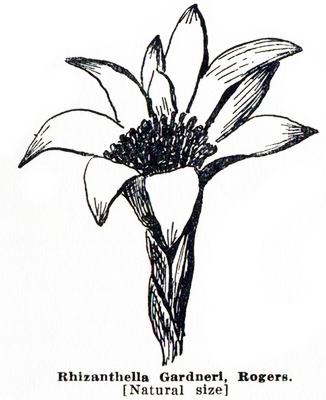
Not actually natural size (anymore).

Most photosynthetic, self-sufficient orchids seem to associate with saprophytic or parasitic fungi. But R. gardneri has found a different niche: myco-heterotrophy, or exploiting the mycorrhizal fungi of other plants. In this case, it gets all up in the business of the fungal partners of Melaleuca uncinata, the broom bush. Mycorrhizae are fungi that encase or invade the roots of nearly every land plant. This is good; without them, plants would have a considerably rougher go of it. The usual exchange is sun-made plant glucose in exchange for a vastly improved water and mineral uptake thanks to the exponential surface area increase made possible by a network of fungal filaments extending well beyond the roots. Mycoheterotrophic plants break into the mycorrhizal associaton of a fungus and another plant and steal some of the goodies for themselves.

R. gardneri is not the first plant to have figured this sweet deal out. Here in Colorado there are at least two pale pink spiky plants terrorize our fungi: the Heath-family pinedrops, and the Orchid-family spotted coralroot. I often see pinedrops and spotted coralroot on hikes in the mountains, where the former associates with (probably parasitizes) mycorrhizal truffle fungi in the genus Rhizopogon (related to the bolete genus Suillus for those who care) and the latter goes after mycorrhizal Russula, which make the majority of the red-capped, white-stalked (and usually poisonous and/or disgusting) mushrooms found in the temperate forest.  I wrote about another Heath-family floral parasite that I used to encounter often here. In spite of exceptions like pinedrops, almost all mycoheterotrophs are orchids.

In all these cases, these plants have lost their chlorophyll. But none have taken the admittedly drastic step (for a plant) of entirely forsaking wind and rain, sun and cloud.

Chloroplasts: Not Just for Photosynthesis Anymore

The Western Underground Orchid has, and in the process, has gone farther toward forsaking its planthood (if its planthood can be tied to the vir(d)ility of its chloroplasts — the cell bits responsible for photosynthesis) than any other plant.

Chloroplasts — as first understood with genius by Lynn Margulis — are what remains of a fateful meal billions of years ago. A bigger cell engulfed a smaller photosynthetic one, probably a cyanobacterium, or blue-green alga. But the alga didn’t go quietly. It managed to survive and thrive in its would-be predator. It became its partner instead. Already in that cell were the remains of bacterial cells who had long ago struck a similar bargain: mitochondria, the power-packs of the cell. You have these ancient bacteria still in nearly every cell of your body. Plants have both mitochondria (which is how we know that endosymbiotic event happened first) and chloroplasts, the descendants of the world’s luckiest alga. And both still have DNA, the remains of their bacterial chromosomes.

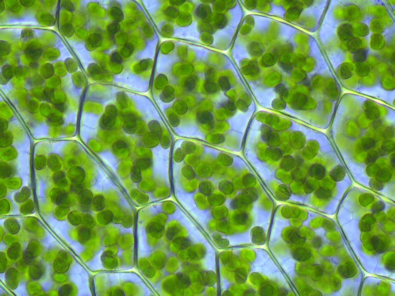
Chloroplasts inside a true moss -- each one a descendant of an alga that gave a predatory cell indigestion billions of years ago. Its offspring (and that of a few other lucky photosynthetic microbes cum organelles) blanket the planet. Creative Commons Krisitian Peters, click image for license link.

But in R. gardneri, 70% of its expected chloroplast genes are gone, according to a new paper in Molecular Biology and Evolution. And the total length of the R. gardneri’s chloroplast chromosome (which is still circular like all most good bacteria) is just 59,190 base pairs, also the smallest for a land plant on record.

Chloroplasts are where the proteins and other cogs of photosynthesis are manufactured. They’re where photosynthesis takes place too. But it seems that even chloroplasts must have some functions in the cell aside from harnessing light. Because Rhizanthella has gone as far as any plant in ditching their chloroplasts since they spend their entire lives underground, ensuring zero photosynthesis takes place. So maybe the news isn’t that they’ve shed 70 % of their chloroplasts genes. It’s that they still have 30% of them. What are those genes still doing there? And what can they tell us about how other parasites operate?

Even in your garden-variety plant, the chloroplast genome is sharply reduced compared to its cyanobacterial ancestor. When a cell goes from going it solo to living in a co-op, there’s a lot of genes it doesn’t pay to make in-house. There may be genes already doing the same thing in the host cell’s nucleus, or just not usable from inside another cell anymore. Over time, natural selection will prune these genes away, since they are costly to make if not being used. Other genes, perhaps because it’s more efficient to regulate and transcribe them there, get shuttled to and permanently housed in the nucleus. What’s left is chiefly what’s essential for the business of turning light into sugar, Job #1 for the choroplast.

But in Rhizanthella, which has clearly boarded up the photosynthetic apparatus, there remain 37 genes coding for 20 proteins, 4 ribosomal RNAs (an important constituent of ribosomes), and 9 transfer RNAs (essential tools for turning RNA into proteins within the ribosome; they ferry the appropriate amino acid to the appropriate RNA codon). Scientists checked DNA made from the messenger RNA they found in the chloroplasts; they were all being spliced and edited correctly, implying they were, in fact, still being used, in spite of the strip mining of the rest of the genome.

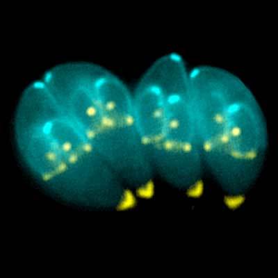
Toxoplasma gondii motherships constructing new invasion craft. Creative Commons Ke Hu and John Murray. Click image for license link.

Compare those 37 genes to a standard photosynthetic orchid, in this case Phalaenopsis aphrodite, whose chloroplast genome has 110 genes. Even the parasite Toxoplasma gondii, notorious for its mind-control abilities (which chiefly involve making hosts — including accidental hosts like humans — do risky things to increase their odds of predation, or from the parasite’s point of view, getting to the next host), actually seems to have been photosynthetic in a previous life, and even it still contains 53 genes on its presumed remnant chloroplast genome, according to the authors.

Thus ex-photosynthetic plants of all sorts seem to share a minimum set of chloroplast genes, and they must be doing so because the chloroplast performs some sort of important non-photosynthetic function that can’t be transferred to the nucleus.

In fact, the scientists compared the reduced gene sets found in parasite ex-chloroplasts from a variety of groups. As mentioned above, they found strong similarities, suggesting that when photosynthetic parasites of any ancestry — related or not — give up free-living, they tend to lose and retain the same sorts of chloroplast genes. This knowledge could provide us with lines of attack in ex-photosythetic parasites that go after us or our crops or livestock. The bare-bones Rhizanthella genes could tell us which genes are most essential — and thus the best targets.

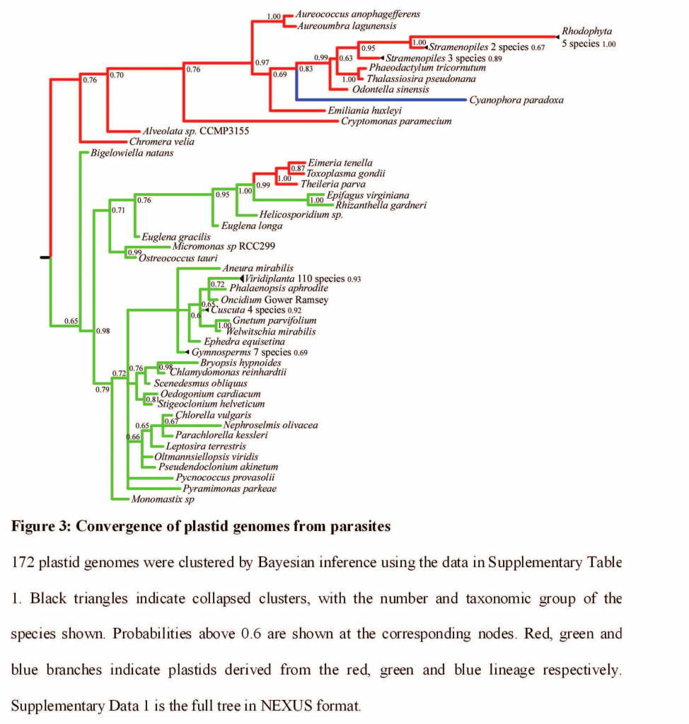
Thanks to convergent evolution, loss of similar genes in parasitic chloroplasts makes it look like Toxoplasma gondii (cause of Toxoplasmosis in humans and cats) is closely related to the plant parasites beechdrops and Western Underground Orchid. Ummm, no.

But wait. Does that mean the chloroplast — the sugar-making machine of plants — does things other than just photosynthesize? Yes. And parasitic plants like R. gardneri are how we know what those things are.

Of the genes that are left on the R. gardneri chloroplast genome, almost all of them encode proteins needed for translation (getting from RNA to protein). Five of the remaining transfer RNAs seem to have remained because either they must interact with many other chloroplast proteins and versions from the main cell would not necessarily recognize the appropriate proteins, or because they have quirky physical modifications that make the chloroplast translation machinery unable to recognize their cellular equivalents. In other words, for these genes, evolution’s backed itself into a corner it’s not easy to get out of with simple chance mutation and natural selection, so instead, it preserves the status quo.

But all these translation-related genes are probably still there for a larger reason: they are necessary to translate the four remaining non-translation related genes on the genome: one making a protein essential for membrane synthesis and two others likely involved in making it, and a fourth gene that makes a protein that seems to have a hand in a variety of important plant processes, at least one of which must be essential. Mutations in the chloroplast’s translation genes are probably fatal because the chloroplast can then no longer make this handful of genes.

Why must these four genes be expressed in the chloroplast and not in the nucleus? The authors hypothesize it has to something to do with a wonderfully vague term called “control by epistasy of synthesis”. As far as I can tell, that means the compartmentalization of these proteins in the chloroplast is essential to their proper synthesis and integration into large protein complexes. So there you have it: the chloroplast may stick around in parasitic plants because it’s handy as a clean manufacturing facility for making parts of a few complicated proteins.

________________________________________________________

* According to the poster of the flickr photograph I used to illustrate this post, Trott donated his specimen to an herbarium for identification and offered £100 to anyone who could find another. Although many looked, none was found until 1979, the year after Trott died. His widow, however, paid up.

ResearchBlogging.orgDelannoy E, Fujii S, des Francs CC, Brundrett M, & Small I (2011). Rampant Gene Loss in the Underground Orchid Rhizanthella gardneri Highlights Evolutionary Constraints on Plastid Genomes. Molecular biology and evolution PMID: 21289370

{ 4 comments }

One organism: three kingdoms. Fnd out what they are, what they look like, and what this entire thing is tonight in Denver.

Tonight I’ll be giving another free lecture, this time at the Colorado Mycological Society meeting at the Denver Botanic Gardens. Doors open at 7:00 and the talk starts at 7:30 p.m.

Here’s the description I wrote:

Take an illustrated journey through the fungi of Colorado. We’ll cover what a fungus is, what the major divisions of fungi are, look at the fascinating microscopic characteristics and other markers of these groups, and admire photographic examples from our own beautiful state.

At the end of this lecture, you should have a better understanding of how a cup fungus and a mushroom are different in a very basic way, and be able to impress your friends with your rust, smut, and bunt knowledge. Whether you’re a complete beginner or an expert looking for a refresher, come get excited for the coming season, and learn how to spot fungi in the field beyond mushrooms!

Hope those of you in the area can make it!

{ 1 comment }

A Protist’s Worst Nightmare

by Jennifer Frazer on March 12, 2011

This video* is hypnotic and illuminating — one might even say joyful, and it is joyful to me to watch it. But if I was a bacterium, alga, or protist (what these rotifers are hoping to get for dinner), I’d feel a bit different. That gauntlet about halfway through the video? Talk about a swirling vortex of rotiferan doom, with their mechanical jaws snapping like clockwork at the bottom of each whirling trap (look for the jaws chugging like pistons about 1/4-1/3 of the way down each gullet).

Repeat after me: “The penitent protist shall pass . . .the penitent protist shall pass . . . ”

As you can see rotifers (literally “wheel bearers”) are so called from the accessories on their foreheads involved in hoovering up dinner. And they are truly amazing creatures. To learn more about bdelloid rotifers (including some gorgeous SEM shots of their jaws) and their alternative lifestyles, see two posts from early in this blog’s life: Lesbian Necrophiliac Bdelloid Rotifers (and the Scientists who Love Them)Parts 1 and 2. The above video provides a good illustration of why they are called “bdelloid”, or leech-like rotifers. They move just like inchworms or leeches**:

Psi Wavefunction did point out, however, that rotifers are *not* the smallest animals. Rotifers are animals (not protists), but as for what the smallest animals might be, I will leave for another day.

_____________________________________________________

*for which I am endebted to the twitter feed of Chris Mah at the Echinoblog

**which are, by the way, annelids (from the last post — see the tree at the bottom) in the Clitellata — look for the Hirudinea. Look for the earthworms just above them in the Lumbricidae. And, just below them but also in the Clitellata (and hence annelids) is the sludge worm Tubifex Tubifex in the Tubificidae — from here, remember?)

{ 7 comments }

The Lowly Peanut Worm and Its Long Lost Relatives

by Jennifer Frazer on March 9, 2011

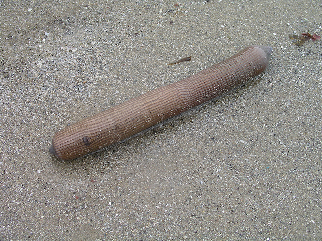
Despite appearances, this is not a naughty present from a bachelorette party. It's a peanut worm from France, with its snout tucked inside. Creative Commons Pgrobe

Breaking news: Peanut worms are annelids. Great! I hear you saying. What’s a peanut worm? (And possible sub-questions: What’s an annelid? And why should I care?) Good questions.

Annelids are the bristled, segmented worms. That is, bristles and segments are the synapomorphies for this group, or the shared, derived characteristics that distinguish them from other groups. A synapomorphy makes their grouping both scientifically valid and evolutionarily based*.

To simplify the classic story: Long ago there was a worm that evolved two special new traits its ancestors lacked:  bristles and segments. This ancestral worm produced offspring that evolved into all the leeches, hydrothermal tubeworms, polychaete bristle worms, and giant Australian earthworms of the world. As descendants of that first innovative worm, they all have segments. And they all have bristles, although sometimes segments or bristles are hard to see — as in earthworms. They also have parapodia, stumpy little extensions of their body on which the bristles are located. And everyone lived happily ever after, until fish hooks were invented. The End.

Well, almost.

Sometimes, the descendants of a creature that develops a distinctive trait may lose this trait for reasons of evolutionary expedience, or simply because there’s no reason not to. These creatures can fool us into thinking they are *not* a member of the group in question. This often happens with parasites that “de-evolve” because they no longer have to make it on their own; I wrote earlier this year about how microsporidia are actually highly evolved fungi called zygomycetes. We earlier thought they were rather bare-bones parasitic microbes, but their loss of many of the zygomycetes’ unique (derived) characteristics initially fooled us. Well, this has apparently also happened to a quirky little group of marine organisms called peanut worms, or “Sipunculids”.
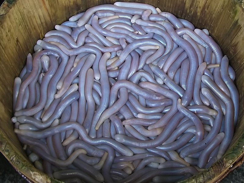
Possible future constituents of "Sipunculid Worm Jelly". As may be guessed, this is a Chinese delicacy. Creative Commons vmenkov. Click for link.

Peanut worms, like so many other strange but real Earth creatures, are Dr. Seussian in design. My favorite observer of biodiversity after David Attenborough, Colin Tudge, describes them thus:

The phylum Sipuncula includes about 320 species of astonishingly unprepossessing creatures, all marine, some roughly resembling sea cucumbers and others like sprouting potatoes, with their tentacled mouths at the end of the sprouts.

Here’s a picture of a potato-esque peanut worm, and here’s a tentacled snout.

They are called peanut worms because when they retract their long snouts (called introverts) and bunch up, they can look like unshelled peanuts. The peanut worm at the top of this post has its snout mostly retracted, and is a bit peanutty. Some peanut worms also have, and I am not making this up, a little calcareous “anal shield”. I am resisting making a crude joke here. There are several I could choose from and I bet you can guess several. I am being strong. Ahem. Like annelids, they also have trochophore larvae, a key character of the lophotrochozoan mother group (the sister of the skin-shedding ecdysozoa in the protostomes, or invertebrates).

Peanut worms have a number of interesting features which you can see drawn out here: a ringed brain, a long nerve cord (like other annelids), and a helical gut that twists back on itself, positioning the anus somewhat near the mouth. Some live in sediment; some crawl into abandoned shells like sand dollars, where they grow so big they can no longer get out; and some, incredibly, bore into rock. Not bad for a tentacled bag.

Like annelids, peanut worms also have a cuticle-coated epidermis, and two layers of muscle — an outer circular layer and an inner longitudinal layer. They have a true coelom (seel-um), or body cavity. Their skeleton acts on hydrostatic pressure, just like an earthworm. Also like annelids, they have a nuchal organ, or ciliated sensory pit, on their head. The mouth at the end of their snout is surrounded by ciliated tentacles and sometimes hooks. Some also have ocelli, or very simple eyes.

We have a few rare fossils of actual sipunculids from the Cambrian(see awesome photos buried in article) — 520 million years ago — and they look remarkably similar to modern peanut worms. Sharks(which have remained similar in for for a mere 420 million years): eat your hearts out. This just goes to show the pace of evolution varies in its own good time. Peanut worms surely evolved during all this time too. They just did so many times slower than uppity groups like vertebrates, because apparently there was little selective pressure to do so.

Amusingly, one of the earliest described species of peanut worm was named Golfingia macintoshii by E. Ray Lankester since he dissected the first specimen provided by a certain Professor Mackintosh between rounds of golf at Saint Andrew’s in Scotland (which for some reason, as an atom in the universe of golf trivia, reminds me of the legendary invention of golf by the Bullroarer Took at the Battle of Greenfields). Golfingia is still the name of a major genus of the organisms.

OK. So now I’ve convinced you (I hope) these things are cool but odd. They share a lot in common with annelids. But also a lot not — no segments. No bristles. No parapodia.

Hmm. Taxonomic impasse. Genes to the rescue!

“I’m gonna need computers. LOTS of computers.”

In a study in Nature released last week, scientists compared the DNA of a variety of peanut worms, spoon worms, and annelids. In this study, scientists used only sequences from expressed genes only. They did this by taking the messenger RNA, an edited form of DNA used to make proteins, and making DNA directly from it (called cDNA, or complementary DNA). They then sequenced this, as DNA pre-edited to make functional proteins is more likley to have meaningful changes to its code due to selective pressure. Bits of non-translated DNA that are edited out are free to mutate much more rapidly and randomly, dampening the signal of evolution.

Scientists can use this method because over time, more closely related organisms will have more similar genetic information. Comparing the sequences of one gene in three creatures to make a simple relatedness tree would be easy for a human, but this study looked at the amino acids (the building blocks of proteins) coded for by the DNA at 47,953 positions in 34 annelid groups. Computers more easily crank through these sorts of hyper-space logic problems, and they did here too. Based on this analysis, the peanut worms fit cozily inside the annelids. When we had only their appearance to go by, we were confused. But with DNA, in this case at least, the situation becomes much clearer.

Sooooooo — to sum up: up until now, scientists suspected peanut worms were in the same branch of bilaterally symmetrical animals as annelids, the lophotrochozoa (who either have crowns of cilia called lophophores or trochophore larvae), but not actually annelids themselves.  You can see where the peanut worms (sipunculids) used to fit in the lophotrochozoans here. Now, scientists see that they’re not just lophotrochozoans, they’re also our old friends the annelids.

Upending the Annelid Family Tree

But the biggest news from their results was not that peanut worms are annelids (although that is good to know), but that our traditional taxonomy of annelids is wrong — and that an old, dusty classification for annelids was right after all. The current system divided all the annelids into the clitellata, or worms that have a collar called the clitellum (that ring you see on earthworms), and the polychaeta, or bristle worms (which, as I’ve written before, have big, showy bristles). But it turns out that clitellata as a group is embedded within the polychaete worms, and that the polychaete worms have non-polychaete worms like peanut worms (which lack bristles, parapodia, or segements, because they lost them) and another non-polychaete that is a weirdo parasite of sea lillies —  the myzostomida, which I wrote about here — embedded within them. That makes polychaeta polyphyletic, which is a dirty, dirty word to taxonomists. It is a group that includes an ancestor and some, but not all of its descendents. Hence, polychaeta’s gotta go.
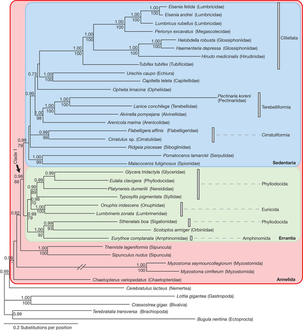
Don’t be frightened by this diagram — it just shows a geneaology of annelids, just like your family tree. The numbers on the tree indicate how certain the scientists/computers feel about their branching hypotheses; numbers close to 1.00 or 100 mean they are very highly confident that that hypothesis of relatedness is correct. And as you can see, with so much data, the scientists are very confident about this tree.

The *new-old* classification supported by the genes was proposed way back in 1865 (though it excluded the earthworms and leeches) by Jean Louis Armand de Quatrefages de Bréau. It is based not on bristling, but on life history — sedentary worms versus active worms. But later scientists dismissed it as the product of another dirty word: convergent evolution — the force that make whales and sharks both seem like they might be a lot more closely related than they are. Quoth the article “This systematization was dismissed in the 1970s as being arbitrary groupings useful only for practical purposes.” Ouch. Well, it turns out that was wrong.

The clitellata(collared/ringed annelids) are still supported as a true monophyletic (ancestor and all its descendants — distinguished by their synampomporphies; taxonomists like these) group. But there are two new groupings: the Sedentaria, which means what it sounds like: things that burrow, or live in tubes, sucking mud or lazily filtering water with tentacles and who could generally could lay around watching re-runs of “The West Wing” for the rest of their lives and not have to worry about their BMI. Things like earthworms, and deep-sea hydrothermal tube worms.

The Errantia are similarly named — they are errant creatures that swim, writhe, and actively pursure their prey or nosh on big algae. Things like the tentaculate and green-bomb-throwing ex-polychaetes I’ve written about here before. On the fringe of these two groups we have some organisms that are neither — like peanut worms — but are still annelids based on their genes and morphology. And there you have it.

Once you have a tree like this, it’s  a fairly simple logic problem to determine what the ancestral form that existed way back on each of those branch points must have looked like. You just look at its descendants and see what they have in common. And here is the drawing the scientists came up with for the ancestral annelid (last common ancestor, scientsts would say) and the ancestral Clitela and Errantia.
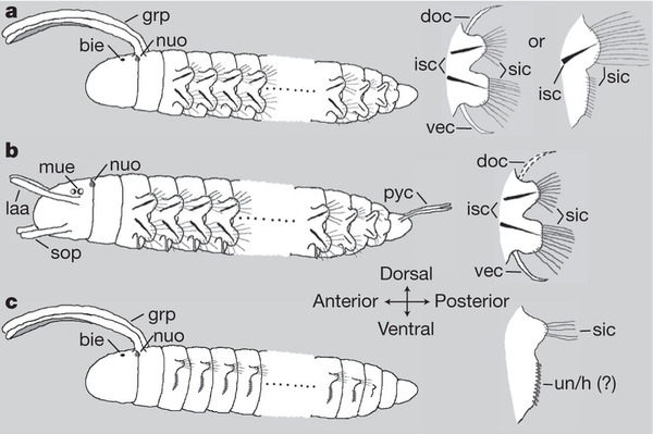
(a) shows features the first annelid likely possessed, (b) the first Errantian, and (c) the first Sedentarian. Note they all have parapodia, though the bristles are much shorter and feebler in Sedentarians, and the parapodia are not supported by stiff internal bristles (chaetae: kee-tee).
As you can see, they have determined that that first annelid worm (a) was indeed segmented, since peanut and another unsegmented annelid I did not cover — the spoon worms, or Echiura — are embedded within segmented annelids, not both at the base of the tree.

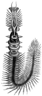
The annelid in the earliest branching position — Chaetopterus, or the parchment worm (see left) — is not only segmented, but quite complicatedly so — it has three different sections of segments on its body.

So it seems the peanut worm simply found that, although segments and bristles were awesome, they were costly options it simply didn’t need to get the job done in its little marine habitats, where it has happily existed more or less as-is, and perturbed by little except predators and Chinese gourmands, for over 500 million years.

———————————————————————–

* Modern taxonomy attempts to base groupings on evolutionary history, since it only happened one way and once. Any other classification system would be arbitrary

ResearchBlogging.org Struck, T., Paul, C., Hill, N., Hartmann, S., Hösel, C., Kube, M., Lieb, B., Meyer, A., Tiedemann, R., Purschke, G., & Bleidorn, C. (2011). Phylogenomic analyses unravel annelid evolution Nature, 471 (7336), 95-98 DOI: 10.1038/nature09864

Arendt, D. (2011). Evolutionary biology: Annelid who’s who Nature, 471 (7336), 44-45 DOI: 10.1038/471044a

{ 7 comments }

Amoeba Tweeting

by Jennifer Frazer on March 8, 2011

UPDATE: Have now placed twitter feed on right sidebar of this blog so you don’t have to go to twitter to see it.

I’ve haven’t announced it here before, but I do have a twittter feed: @JenniferFrazer. Lately I’ve been trying to use it more as a way of sharing links and ideas that I don’t think warrant full posts or don’t fall within the purview of my blog per se, but that are still way cool. If you tweet, consider following me!

Full blog post upcoming tonight on one of the many, many suggestive-but-obscure worm groups — promise. : )

Ed. note — lost internet access until late. Post TK early a.m.

{ 3 comments }

Empire of Light

by Jennifer Frazer on March 3, 2011

“His only hope for existence on this planet is as a gigolo. He’s gotta find himself a babe, and then he’s gotta latch on for life.”

I missed Edith Widder’s TED talk on ocean bioluminescence when it came out in 2010. Had I known, you would have been seeing this much sooner. It clocks in at 17 minutes, but don’t let that intimidate you; it speeds by. There’s a little something here for the biophiles, engineers, and daring explorers in all of us.

For a high-res version of this video, click here.

What I would have given to have been in that Wasp suit when she turned the lights out.

That jellyfish burglar alarm is just fantastically beautiful. Notice how the pattern is slightly different with each rotation. Why does it remind me of an ’80s electronic toy?

For the record, I am so with Edith on the whole, “When someone offers you a trip to the deep sea, you say YES.” In fact, I’m actively looking for a low-budget ticket to several thousand clicks down; it’s a dream of mine. I’m no Dennis Tito, but I can write. If you know someone traveling to the deep sea who might be in need of a science writer specializing in biodiversity and natural history who doesn’t eat much and fits conveniently in most overhead bins (5’2″, 108 lb.), LET ME KNOW.  : )

For a more in-depth look at deep sea life which Dr. Widder helped work on, see the Deep Sea episode of Blue Planet, available through netflix in the US (and mercifully narrated by David Attenborough, and not Oprah). You will not regret it.

{ 1 comment }