Though we don’t think about it very often, there’s a universe of amazing life humming along inside you. For example, inside nearly every living non-bacterial cell in your body, you will find …
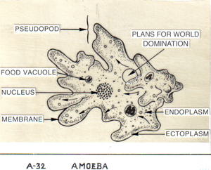
This remarkable video is one of a series recently produced by a biological animation initiative at Harvard. Earlier, they made a more extensive movie of the inner workings of a cell (See the adventures of a white blood cell in The Inner Life of the Cell here, and the considerably less inspirational but more enlightening narrative explanation of the video here (this is what happens when science writers do not write the script)). They’re not new; Carl Zimmer has posted one of these videos before, but I wanted to make sure none of you missed them. They’re a far cry from the cartoonish still drawings (see below) we had to use our imagination to envision in real life when I was in school.
So what’s actually going on in the mitochondrial video? Well, I’m not sure of every detail, but as a former biochemistry TA, I can give you a lot of educated guesses. The movie appears to begin with mitochondria — sub-cellcular power stations — inching along a part of the cellular skeleton called microtubules. The job of mitochondria is to finish the process of turning the energy stored in the bonds of glucose into useable power through respiration. As energy is harvested from the electrons prised off glucose, they are finally passed to oxgen, which accepts them along with some hydrogen ions to form water. Meanwhile, the carbon that was tied up in glucose ends up as carbon dioxide. This forms the basis of your inhalation of oxygen and exhalation of carbon dioxide with each breath you take. Anyway, back to our film.
Then an (?)amino acid chain (fragment of a protein) of some sort escorted by (?)chaperone proteins enters a mitochondrion via a pore through its double membranes*. Then you see the contents of the mitochondrion: an asteroid field of colorful proteins zipping about amid strands of the mitochondrion’s DNA.
Then, looming in the distance like the Pillars of Hercules (or the warship encrusted columns inside the motherships in Independence Day — which seems to be the point. They are going for cinematic here.) are tubular mitochondrial cristae, or folds of the inner membrane. These folds increase the surface area available for respiration.
Embedded in these columns are the rotary engines of mitochondria — enzymes called ATP synthases. These proteins are fascinating feats of natural selection that rotate as they charge their substrates (the molecules they will act upon): namely, ADP. Swarming around these proteins like fireflies are hydrogen ions (H+ — essentially, a proton, but often surrounded by three oxygens in aqueous solutions) generated by stripping glucose of electrons like a car in chop shop.
Without going too deep into the gory details, when a cell burns glucose, it performs some preliminary reactions in the cytoplasm (glycolysis) and sends the remaining energy-bearing bits into the mitochondrion for full processing. After enzymes performing the Citric Acid cycle in the interior of the mitochondrion (called the matrix) squeeze more power out and release what’s left of the glucose as carbon dioxide (CO2), the electrons glucose has yielded are passed down the electron transport chain of proteins embedded in the inner membrane, which use the energy thus released to pump hydrogen ions out of the matrix into the space between the mitochondrion’s two membranes. I think you can see this happening at about 1:08.
The inner membrane, unlike the outer, is highly impermeable to most molecules — even to tiny hydrogen ions. The resulting ionic gradient can only flow back downhill through a rotating pore in ATP synthases**. The passing hydrogen ions powers their rotation and their charging of ADP to ATP, the cell’s energy currency (an explanation of this fascinating mechanical process can be found here under “binding change mechanism”). It’s such a clever system that engineers have designed engines (called the Wankel Engine) based on the same principle and built them into working cars — namely, the Mazda RX-7 and RX-8. I know this because one of my biochemistry professors at Cornell had actually owned one of these for that very reason (You know you are a nerd when . . . ).You can see the biological version of this process happening at about 1:15, where small molecules enter the head of the synthase, light up to let you know they’ve been charged, and then are released to please go play nicely with the rest of the cell.
Here is a conventional representation of what I just described — I, II, III, and IV are proteins of the electron transport chain, NADH is an electron ferry that shuttles said particles from ex-glucose pieces to the electron transport chain, and succinate is an intermediary in the citric acid cycle (for those that remember, this is the step that generates FADH2):
![]()
During all this action in the movie, the camera also passes once or twice through the undulating lipid bilayer of membranes, where the kinky double-tailed (and faintely spermish) phospholipids jostle against each other to keep the membrane fluid. Mitochondrial membranes actually contain many fewer sterols (cholesterol is one — they are molecules that help stabilize membranes) than the cell membrane, giving the mitochondrion greater shape-shifting powers.
I think the next-to-last scene is an ATP/ADP transport protein that actively shuttles ADP into the matrix and ATP out. Finally, you see all the mitochondria swarming toward some big shiny thing (centrosome? endoplasmic reticulum? Who knows! Scene list please, Harvard!) like star cruisers converging on a galactic rendezvous. Actually, mitochondria do sometimes cluster near where they are most needed. For example, in cells with flagella, they may cluster near the base of the tail.
All in all, the mitochondrion’s a tightly run ship. Lest the ID community use these incredible little machines as evidence of “stasis” or “irreducible complexity”, let it be known that anaerobic (non-oxygen breathing and non-mitochondrial) bacteria alive today have proteins almost identical to ATP synthase that function in reverse: powered by ATP, they serve to detox the bacteria of H+ to rid them of the acidic by-products of the less-efficient but still-better-than-nothing energy-producing process of fermentation. The cytochrome complexes (aka I, II, III, and IV, the cogs of the electron transport chain) may have evolved for similar detox purposes in other ancient bacteria before being combined with ancient ATP synthase by natural selection to form the well-greased respiratory engines we have today.
Typical plant and animal cells contain hundreds or thousands of such mitochondria, though their number ranges from one bizarro giant in a few single-celled protists to several hundred thousand in well-provisioned egg cells. I’m not certain why the directors of this film chose to show this mitochondrion with tubular cristae. Most vertebrates have regular laminar, or sheetlike, cristae (remember that it was unusual that alvaeolates (the paramecia, ciliates, dinoflagellates, and apicomplexans) had tubular cristae), though plants have both sheets and tubes in their mitochondria, and are more irregularly shaped and sized.
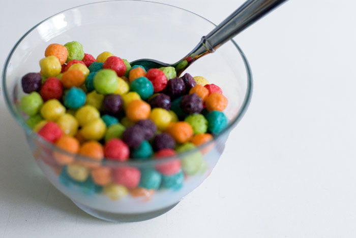
Of course, proteins inside mitochondria don’t really float around looking like someone blew up a box of Trix in the space station. In reality, my understanding is they’re all sort of, well, clearish at that scale. And in this thorough NYT article on recent advances in molecular animation, scientists acknowledge that molecular animators also take liberties with space.
“Some animations are clearly more Hollywood than useful display,” says Peter Walter, a Howard Hughes Medical Institute investigator at the University of California, San Francisco. “It can become hard to distinguish between what is data and what is fantasy.”
But clearish molecules and vast distances would make for a pretty dull movie, so I don’t begrudge them their colors. This situation reminds me of the Peter Jackson Conundrum: was the Lord of the Rings better before your head was filled with Peter Jackson’s version of everything? And wasn’t it better when only the people who actually took the trouble to do all the reading were in on the magic?
My gut feeling is that these movies are a good thing, as is sharing the wonder with the masses. If we wish to make the case to society that science is important and worthy of time and money even on its own terms, animations like these help. It is also unquestionably cool to see it all in such detail — revealing things we could not easily foresee without seeing everything together in glorious living color — even if our imaginations are a bit impoverished for it. It’s a worthy sacrifice, in my opinion, if our appetites are whetted.
________________________________________________
* The outer membrane is a product of a long-ago engulfment of a bacteria by a predatory cell — the inner membrane is the ancient bacterial membrane and the outer membrane is the erstwhile vacuole. Further evidence for endosymbiosis includes that mitochondria (and chloroplasts) have their own single, circular (like most bacteria) DNA-based chromosome without a nuclear membrane from which they manufacture their own proteins and bacterial-sized ribosomes (which can even be interchanged with bacterial ribosomes in some cases) and replicate by division. They’re bacterially-sized too: 1.5 by 2-8 micrometers.
** In high-magnification photos of mitochondria, you can actually see the ATP synthases poking into the matrix like lollipops.
{ 1 trackback }
{ 0 comments… add one now }