When Ostracods Fly and Copepods Fire

by Jennifer Frazer on September 22, 2009

I’ve been treating myself to the BBC’s Blue Planet: The Deep this week, and there are delights in every second of film. One that particularly caught my interest is below. Those of you who recall my post on Swima bombviridis will recognize the same strategy, if not the same artillery, at work here.

Now you have to admit — that orange ostracod is even more amazing than the depth-charge wielding copepod. I mean come on! Had you ever imagined a creature like that could exist? Most ostracods are considerably smaller and are called seed shrimp because of their resemblance to something like a bell pepper seed. The males should get some sort of award for virility; many species have two penises and sperm that can be up to six times the male’s body length when fully uncoiled. The ostracod in this film appears to be  a female, given the clutch of round objects residing in its posterior.

The filmmakers here seem to subscribe to the Star Wars school of foley-artistry: no sound in space? No problem! We’ll just give the spaceships cool sounds anyway (good call). Bioluminescence is silent? No problem! We’ll just give flashing creatures Super Mario-grade sound effects . . .  No offense meant, though, Blue Planet dudes at BBC. I worship your work. Unlike Macgillivray Freeman, who in my opinion have largely squandered the IMAX format with their insultingly stupid and poorly written scripts, in spite of brilliant cinematography . . .

Annnnyway . . . .Copepods and Ostracods are actually fairly closely related, both being Crustaceans. Check out the copepod taxon here, but make sure to check out the next highest taxon, Crustacea, to see how copepods and ostracods fit into it. Jumping down the ostracod hole will take you here.

{ 2 comments }

Does This Membrane Clash With My RNA?

by Jennifer Frazer on September 17, 2009

Does this membrane clash with my chromatin? Image courtesy CDC/ Erskine. L. Palmer, Ph.D.; M. L. Martin

A membrane, some proteins, and 8 segments of RNA: all there is to influenza. A false color transmission electrion micrograph of the influenza virus. Image courtesy CDC/ Erskine. L. Palmer, Ph.D.; M. L. Martin

Until 1933, it was impossible to see a virus. Oh, we knew they were out there. But no one had the faintest clue what they looked like. 1933 marked the year transmission electron microscopy finally achieved resolutions finer than light microscopes were capable of and made it possible to finally glimpse the agents that had mottled tobacco leaves, streaked tulip petals, scarred the faces and bodies of millions, or paralyzed, maimed, and killed millions more.

So what’s with the doofy colors? Yes, in spite of the awesomely awesome resolution that transmission and scanning electron microscopes provide us with, scientists and alarmist pandemic book cover designers can’t seem to resist painting them with gaudy colors (see above). OK, I admit the colors do seem to spice up the images. But this isn’t even a case of colorizing something that was colorful to start with — viruses are quite clear. So what a revelation to see a glass artist team with scientists to produce anatomically correct transparent glass sculptures of viruses and other wee animalcules. That’s exactly what British artist Luke Jerram has done, and his creations are truly illuminating.

Anacardium occidentale -- hard to believe, cashews actually *do* grow on trees.

It's hard to believe, but cashews actually *do* grow on trees. A glass model of Anacardium occidentale, the cashew tree, on display in the Harvard Glass Flower Collection. Model by Leopold and Rudolf Blaschka.

I must say that his models remind me very much of the intricate glass 19th century models of fungi, invertebrates, and plants I discovered in my college days in dusty corners of Cornell and Harvard, many of which were created by the father and son team of Leopold and Rudolf Blaschka in Dresden, Germany from the 1880s to 1930s. They definitely get my vote for having the C00Lest Jobs EVAR. Intended as teaching aids, they date from a time when color photographs were unheard of and microscopes were a bit primitive. The colored glass models were able to show fine detail far better than either an engraved image or tiny eyepiece could, they did so in 3D, and as the Harvard people like to point out, glass flowers bloom year round. It’s nice to see that everything old is new again.

Viral family trees

{ 0 comments }

Tree Time

by Jennifer Frazer on September 15, 2009

An acient tree with a bit of a bias. The true tree of life is trunkless -- more of a shrub of life, really.

An 1879 tree by Ernst Haeckel with a bit of a bias. The true tree of life is trunkless -- more of a shrub of life, really. There's a root, but no apex. If forced to choose one, personally, my money's on Theobroma cacao.

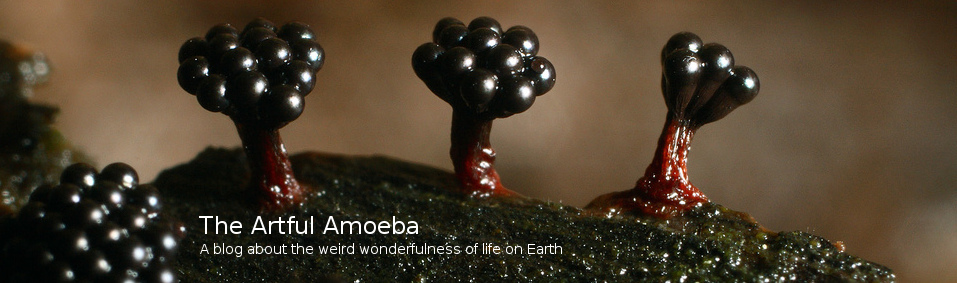
Last time I posted a link to a slide show of beautiful jellies. But I don’t want this blog to be only about eye candy. I want to help you learn about new organisms, the often crazy or amazing ways they make their livings, and no less importantly, how they are related and classified.

Because I hope to make this blog accessible to all sorts of readers, from precocious 10-year-olds on up, I’ve struggled with how to help you learn about taxonomy without making you digest the long lists of incomprehensible names found in abundance on most trees. On top of that, I face the problem that classifications are constantly changing.

The Trouble with Trees

Today scientists classify organisms based on how they are related to one another, but unfortunately, it’s often quite confusing to figure out. Sometimes comparing one trait — say, tentacle length — yields  one family tree (often called phylogenetic trees by scientists), and comparing another trait  — say, mean number of biologists devoured attempting to study organism — yields a conflicting one. Which is correct? Which traits should you give more weight when constructing the tree you think most likely? This is the problem that has launched a thousand theses. Scientists argue about the true relationships constantly, and the trees are rearranged with every publication of a systematics journal.

On top of that, once scientists started sequencing the genes of different organisms and making trees by comparing them, traditional taxonomies that had been stable for decades or centuries based on body shape, anatomy, or other observable traits were often upended, leaving things in disarray to this day. And finally, the formal names we give ranks above species like kingdom, phylum, class, order, etc., are largely arbitrary, as is the idea that there are exactly seven ranks. There aren’t. The ranks are meaningless as absolute markers, so teaching these names seems to me both confusing and pointless.

And yet . . .

The Learning Tree

Some major groups have remained supported by scientific consensus, and other new groups are settling down. And there are true evolutionary relationships among organisms, and themes within lineages of common descent, though individual species can differ radically from their close kin. Learning the major groups helps keep the dizzying diversity of Earth organized in our brains. Strange new species will no longer float around like stray mental post-it notes, but have a taxonomic hook to hang on when you can say . . . ah, that new creature is an annelid. I know exactly which other creatures it’s related to.

So I’m going to try to start including links to trees with each post. It’ll be up to you to explore them as your fancy strikes you. One site I will rely on heavily is the Tree of Life Web Project. Although the descriptions there are often written by scientists for scientists and will be nigh incomprehensible to the lay person, anyone can look at the trees and get a sense of who is related to who and how. Plus pretty pictures help with scary names. : ) Another benefit  to studying these trees is seeing how many different organisms are out there that you will never have heard of, and about which so little is known. Virtually every page contains groups that even I — with six years of higher education in biology and a passion for, shall we say, creative life forms — have never heard of.

So here we go: For jellies and friends, which are contained in a group with the formidable name Cnidaria (ni-DAR-ee-a), you can see the TOL trees here and here and a cuter and more digestible, if less rigorous, tree here. Cnidaria was one of the first animal groups surviving today to split from the rest of the animals — and it shows.

{ 0 comments }

Juxtaposition of Jellyfish

by Jennifer Frazer on September 10, 2009

Come here often?

"Come here often?"

Here in Boulder there is a restaurant that serves the proposterously-named “Juxtaposition of Duck”. I could not resist when it came to titling this post. From the Beeb, I present to you a gorgeous eye-candy gallery of Arctic jellyfish. One of my great delights in studying the diversity of life on this planet is the variety of form, texture, and color. Slime molds, lichens, and jellyfish, in particular, provide some of the best highs. Enjoy, and Happy Friday.

BTW, is it just me, or does it seem like the caption for #2 should be, “Luke, I am your father!”?

{ 2 comments }

Slime mold sporangia (spore capsules) broken open and laid bare to the wind as nature intended. A few unopened capsules are on the left. Photo courtesy Mary Jane Howell.

Slime mold sporangia (spore capsules) broken open and laid bare to the wind as nature intended. A few unopened capsules are on the left. Photo courtesy Mary Jane Howell.

At last, two actual slime molds! As hard as I searched, the best I could find were plenty of tiny white-spored objects (fungi) erupting from the wood. I learned slime mold spores tend not to be white. : ( But local slime mold expert Mary Jane Howell’s sharp eyes picked out two (not so slimy) slimes. One, a Stemonitis, often called “hair growing on wood” because of their long spore-making sacs called sporangia, had lost most of its spores and was fairly unexciting. Only the hairnet-like frame of the sporangium was left, and even that was a bit disarrayed.

20090831_0272a_5
But another, Perichaena corticalis (top photo), still showed the bottom half of its spherical spore capsules and a thin veneer of yellow spore dust.

I also learned there is a certain age of fallen log that is ideal for slimes — bark starting to come off, but not all off, and that looking underneath the log or bark can also bear fruit. To review, plasmodial slime molds start out as spores that hatch into amoebae or flagellated swimming swarm cells. The amoebae crawl around for a while feeding on bacteria and other microorganisms in the soil until they run into a mate.

After they fuse, they eventually start pumping out nuclei and more cytoplasm like crazy but everything stays in one big bag — the plasmodium — that crawls around until things dry out. They then produce the fruiting body, of which the spheres  of Perichaena above are one form.  When those dry out and crack open, the spores are distributed by the wind.

Fungi and slime molds aren’t the only thing I look for when I’m out. I also pay attention to lichens and plants, which have the indisputable advantage of being much more abundant and much less dependent on recent rain for viewing. I found a new (to me) species of bog or rein orchid (Platanthera sp.), pixie sticks and pixie cups (lichens, probably Cladonia sp.), and everywhere the jewel-like fiery red pendant berries of clasped-leaved twisted-stalk (Streptopus amplexifolius) of the lily family. Lilies are monocots, one of two major flowering plant divisions. These plants tend to have parallel veins and flower parts in threes — if you click on the link, notice the leaves with parallel veins and 6-petaled (tepaled, actually) flowers hanging from their twisted stalks.

Finally, we found, in turning over damp branches, several of these tiny snails. As you can see, they are plenty slimy, and plenty cute.

Slime Mold Hunt Aug 2009 001

20090830_0249b_1

Do we have any invert experts here who can ID them? Here ends the tale of the slime mold hunt!

{ 1 comment }

cropped_rust
When I was young, I used to think that leaves just naturally got spotty and brown as they aged. But after taking plant pathology, I learned those spots you seen on leaves are almost always fungi or insects infesting plants, and careful examination with a hand lens can reveal a whole new little world to you. As we walked through the woods last week, I saw many leaves with yellowing spots on top. I turned them over, and lo, underneath were creamsicle-orange piles of rust spores.

Rusts are one of the oldest known plant pathogens, and the Romans even had a god –Robigus — dedicated to staving off the stuff (and that included the rust of metal — not having microscopes, Romans considered them one in the same). Rusts are fungi that go through incredible reproductive gymnastics, producing up to five seqeuential spore types in five different reproductive structures and jumping among two or three different hosts per life cycle. I think the rusts are abundant this year because we had such a wet June in Colorado, but that is just a guess.

Heavy infestations hurt plants, but in the woods they are very pretty to see if you turn over leaves to see the eponymous rusty-orange urediospores underneath. Mycologists have long, difficult-to-pronounce names for each rust spore type and spore-making body (naturally), but we’ll save those for another post. Just like our jelly fungi and zygomated mushroom, rusts are basidiomycetes, one of the four major groups of fungi. The others, for reference, are chytridiomycetes, zygomycetes (as we saw last time), and ascomycetes.

Ascomycetes are fungi that make their spores inside sacks called asci (yes, pronounced ass-eye (singular ascus: ask-us). My plant path grad school department once had a soccer team named after asci that grow without an enclosing structure: “The Naked Asci”), and we found several of these on wet wood too. Many, but by no means all, come in cup form. Eyelash cups (Scutellinia scutellata), in particular, peppered many waterlogged branches with their lashed but lidless eyes.

A body double for our Molly eye-winkers -- I forgot to get a picture. Image courtesy Dan Molter, Creative Commons 3.0 Attribution-ShareAlike 3.0 license. Click image for link.

A body double for our Molly eye-winkers -- I forgot to get a picture. Image courtesy Dan Molter, Creative Commons 3.0 Attribution-ShareAlike 3.0 license. Click image for link.

Next time: Slime molds. I promise. Also snails. Cute snails. You know you can’t resist.

{ 0 comments }

Not what you're thinking, I swear . . .

Not what you're thinking, I swear . . . Photo courtesy Mary Jane Howell.

Well, the big slime hunters (slime mold expert Mary Jane Howell and I) have returned from our foray but, alas! without many slime molds. Nonetheless it was a day well spent, full of fluttering green aspen, mossy corners, and bubbling pools. We crawled our way into the moistest, juiciest spots we could find, but unfortunately, in August in Colorado in a year without a proper monsoon (as our traditional early August rains are known), it’s crispy out there. The bogs are boggy and buggy, but the logs are dry on top and the slime spores have flown.

What you see in the picture above is not a slime mold. It is a jelly fungus, called so because of its appearance and slick, rubbery texture. If you ever see one of these, feel free to reach down and pet it — they feel delightful and do not bite or sting. Jelly fungi are basidiomycetes like mushrooms. That means they make their spores on the outside of special reproductive cells called basidia (singular: basidium).

Then there was this:

20090830_0255a_3

A pincushion? A voodoo mushroom? A balding mushroom that decided to get "plugs"? Photo courtesy Mary Jane Howell.

Super cool! This is an LBM (little brown mushroom) of some sort that seems to be getting parasitized by a zygomycete (like Rhizopus stolonifer, the common bread mold). Zygomycetes in the order Mucorales have a characteristic asexual spore structure (a sporangium) that often looks like a black ball at the end of a long filament, as you see here. There were several mushrooms in the group that had fallen prey, and a few that so far had escaped.

Up next time: More slimy, rusty, and snaily goodness from the woods of Colorado.

{ 4 comments }

Gone Slime Mold Huntin'

by Jennifer Frazer on August 30, 2009

aug-oct2008-005

In yet another sign of what an incredibly big dork I am, I have arranged to go slime mold collecting with Rocky Mountain National Park’s volunteer slime mold expert. We will be going to an undisclosed mountainous location well outside the park. I am bringing my shockingly inadequate camera but if I find anything worth sharing, I will definitely post it here later this week! I need hardly add that it is still bolete and chanterelle season, so with any luck, I will have something for both brain and table. Happy Sunday . . .

{ 0 comments }

Two Sides to Every Worm: the Polychaete Nereis

by Jennifer Frazer on August 29, 2009

In honor of our foray into the world of polychaete worms this week, I thought I’d share some closeups of a polychaete that has been the subject of many an invertebrate lab dissection: Nereis.

In this first video you can see our subject looking kind of cute and shy (awwww!). Pay attention to the dorsal (top) vessel and you’ll see the “human-like blood” being pumped through the worm’s closed circulatory system (just like us!). In Nereis, the dorsal vessel itself does most of the pumping. Also note double eye spots (this guy is literally a four-eyes) and the leg-like parapodia with their bristly setae.

But in this video you’ll find Nereis has a second, distinctly not cute side: vicious predator. Watch for it to evert its pharynx, and particularly watch for the TWO GIANT FANGS (technical term: jaws) on the end. You can also see the pumping blood at the beginning of this film. Unfortunately, this one is a bit dark.

Although you may be tempted to think these guys are closely related to millipedes and centipedes, they’re not. This is yet another case of  — you guessed it — convergent evolution.

{ 1 comment }

Have You Seen This Creature?

by Jennifer Frazer on August 25, 2009

‘Cause scientists sure haven’t. And they really, really want to. The creature in question is Paleodictyon nodosum. And before you do anything else, go check out this article in the New York Times by William J. Broad and take a gander at it. If this is a blog about the weird wonderfulness of life on Earth, I don’t know how something could qualify more. Whatever this is, it is very weird, and it is very wonderful.

DSV Alvin sets a lander basket with tube cores on the bottom. Credit: National Oceanic and Atmospheric Administration/Department of Commerce

The sort of habitat our mystery creature lives in, and the submersible that has tried to find it. DSV Alvin sets a lander basket with tube cores on the bottom. Note the encroaching darkness. Think of yourself living in that environment -- a soft mud bottom, and nothing but miles and miles of cold, inky blackness, as far as the eye can't see. Credit: National Oceanic and Atmospheric Administration/Department of Commerce

There are a few creatures on Earth we knew as fossils before we met face to face. Take the coelacanth. Scientists were shocked to discover a very much alive specimen of this be-lobe-finned fish hauled from the depths off South Africa in 1938. Prior to the discovery of this bit of rather irrefutable evidence, scientists believed the fish went the way of the dinosaurs (literally) at the end of the Cretaceous, 65 million years prior. Although not the first, Paleodictyon is probably the only member of this fossils-first group that was briefly considered to be evidence of some sort of alien deep sea race (hellooooo, Abyss) before it was connected to its fossil ancestors, essentially unchanged after 500 million years.

According to the article, scientists have suggested the hexagonal tubes they have found may be bacteria farms, worm burrows (or both), or the trace fossils of decayed compressed sponges that have long ago been scavanged. The paper even suggests such a sponge may have ties to the Ediacaran fauna, a class of bizarre creatures that preceeded the Cambrian Explosion. There’s one other candidate for Paleodictyon‘s identiy: a xenophyophore. They are the subject for another blog post, but the short, short version is that they’re gigantic single-celled organisms big enough to fit in the palm of your hand, which (like slime molds!) are multinucleate and feed by engulfment using pseudopodia, and (unlike slime molds) inhabit casings they put together with odd things lying around, including (sometimes) their own feces. In spite of being startlingly obscure, these things are apparently quite abundant on certain parts of the ocean floor. Still, this possibility doesn’t quite seem to fit the bill as no xenophyophore crunchy bits have ever been found in the hexagonal holes.

What about you, readers? What do you think Paleodictyon nodosum is? If you think you know the answer, write it on the side of a Deep Flight Super Falcon High Performance Winged Submersible with carbon fiber pressure hull, dual cockpit flight controls, heads-up instrumentation, and laser “collision avoidance feeler beams”, and mail it to Jennifer Frazer, General Delivery, Boulder, CO 80301. Or put it in the comments below (boooo-ring!). Creative answers encouraged!

{ 2 comments }