Mind the Rock Snot

by Jennifer Frazer on August 17, 2010

Creative Commons Thorney¿?

Fly fisher(wo)men everywhere are mourning the loss of a cherished piece of equipment: their felt-soled waders. All too often now, clinging to the felt fibers are the tenacious strands of Didymosphenia geminata (did-em-o-sfeen’-ee-a jem-i-na’-ta), or, for the rest of us, rock snot.

The stuff looks like pre-owned toilet paper and apparently feels like wet cotton, and it’s slowly taking over the freshwater streams of the temperate world, smothering fish, insects, and other aquatic life. It spreads by hitchhiking on the gear of flyfishers, challenging slime molds, dandelions, and jellyfish for the non-human Plans for World Domination Cup. You can read all the gory details in New York Times articles here and here. But hidden inside that slimy brown mass is a work of remarkable beauty.

This.

With the lines of a Stradivarius and the detailing of a Fabergé egg, this baby is a microscopic work of art. If only its macroscopic manifestations could be so beautiful. As you may have guessed, it is a diatom (as covered here), a microscopic glass house (literally (littorally?) made of silicon dioxide) enclosing a little photosynthesizing alga.

At left you see two interesting features: The two long slits, or raphes, through which the diatom can secrete mucilage (aka slime) with which it slides over surfaces, and the porefield, through which it can secrete a mucopolysaccharide (aka slime) stalk that attaches it to a surface. The secretion and aggregation of these stalks is what causes the brown mess of rock snot, not the beautiful fiddle-like head.

In beauty, destruction. In destruction, beauty. This particular destruction brought to you by the otherwise largely upstanding diatoms, conveniently located in this sector of the tree of life. For all the scientific, er, dirt, on rock snot, including a beautiful scanning electron micrograph of the trouble-causing stalks, check out this EPA White Paper.

{ 10 comments }

A Fungal Family Album

by Jennifer Frazer on August 16, 2010

This object is one of my mortal enemies -- and one of the most beautiful fungi I know. Creative commons vinconter

Oops! Note corrected time below. But come early to see the mushrooms that will be on display on the tables!

For those of you in the Denver/Boulder area, I’ll be giving a short free lecture tonight at the Colorado Mycological Society meeting on fungal biodiversity called “A Fungal Family Album” in honor of the International Year of Biodiversity. It’ll be like this blog, but with me standing in front of the screen talking. Plus you can come see lots of fresh mushroom specimens before the meeting!

7 7:30p.m. at The Sanctuary Downtown (formally – last year – the Asbury Event Center) at  3011 Vallejo Street in historic North Denver (map).

{ 0 comments }

So, Twinflower, We Meet At Last . . .

by Jennifer Frazer on August 11, 2010

An event of tremendous biological import took place in my life last weekend. And no, it wasn’t the 300 lb. black bear that wandered through our camp (although that was of tremendous import in that particular moment). No, an event I’ve been hoping and waiting for for many long years took place. I finally found the twinflower, Linnaea borealis.

Examine the buttonhole.

I have been looking for this flower for years, not least because it was Linnaeus’s (as in Carolus “Father of Taxonomy” Linnaeus) favorite flower in the world. Nearly every painting you see of him shows him clasping or otherwise displaying a pair of the dainty blossoms. They were on his coat of arms.

For years I’ve gazed at them in my flower books, hoping and waiting and trying to be patient for the day. That day was Saturday. My mushrooming buddy Johnny was there to see it, and he patiently endured five minutes of me exclaiming over the low mat of little pink flowers. I tried to sniff for the “light vanilla scent” one of my books advised me they would have, but I could detect nothing. I don’t care. They are awesome.

Twinflowers are in the Honeysuckle family, the Caprifoliaceae (Kap’-ri-fo-lee-ase’-ee-ay). The Honeysuckle family is notorious for producing flowers in . . . you guessed it . . . pairs. The sweet honeysuckle blossoms of southeast Tennessee I remember from my youth came in pairs; the kids used to say you could pluck them and suck the nectar, though I don’t recall ever being successful at that. At the foray this weekend, someone came up to me to ask me about another plant he’d found with glossy twin black berries mounted on shiny red bracts; it was the bracted honeysuckle, or black twinberry, yet another member of the family. It was a Caprifoliaceae kind of weekend.

Twinflower is unusual because it grows in a low green mat rather than a woody shrub, like most honeysuckle. I even found a clump this weekend growing right on top of a tree stump (pictured above. You can also see the pixie stick form of Cladonia lichen mingling with the twinflower as if they were at a cross-kingdom cocktail party). Twinflower is, as its name implies, circumpolar in the northern hemisphere, which is why both Linnaeus and I can enjoy them, despite the fact that I’ve never been to Sweden, and he never experienced the Rocky Mountain High.

{ 2 comments }

Why Is My Snow Bank Moldy?

by Jennifer Frazer on August 11, 2010

Find OUT why in my first freelance magazine article over at High Country News: The Drift Dweller. Hooray! I’m a journalist once more.

{ 2 comments }

A Stinkhorn in Need of Viagra

by Jennifer Frazer on August 6, 2010

You may have noticed I’ve been a bit more . . . MIA than usual. That’s because it’s summer and I’ve been out in the field! One of my latest finds, er . . . popped up this week in very nearly my own back yard: my first-ever stinkhorn. Yes, it took me 32 long years to finally catch one in flagrante delicto.

Sad Stinkhorn. If only it had access to the little blue pill!

I covered a new species of African Lacy Stinkhorn in a post here before, but let’s face it: there’s no such thing as too many stinkhorn posts. For those of you who need a refresher, stinkhorns are fungi that hatch from “eggs” enveloped by a peridium (you can see the remains of the peridium at the tip of the head, or receptacle, and at the base of the stalk, above). Some brave souls profess to enjoy eating the eggs. Once the mushrooms hatch and reveal that they are, in fact, quite happy to see the world, they spread their spores by giving them that special odeur de corpse, thereby attracting flies who do the two-step in the sticky, slimy mass of spores at the tip. The flies eat some of the mess; some of the rest clings to their feet. When the flies land elsewhere (i.e. nearby soil, a garbage can, or your sleeping forehead) the spores are deposited  in a new, hopefully stinkhorn-friendly place. Then the spore germinates, and microscopic filaments called hyphae spread out through the soil to . . . I’m not exactly sure what. The one thing I cannot discover is whether stinkhorn fungi are wood rotters or symbionts (partners) with the roots of trees or other plants, the two chief fungal m.o.s.

They’re in the same general group as the gilled mushrooms, but in a special family all their own called the Phallaceae. (Fal-ace’-ee-ay) Some of their brethren are among the most striking fungi on the planet: the earth stars, earth cages, and the lacy stinkhorns, which have a demure, delicate skirt jarringly draped around the obscene fungus. This group, in turn, is in the Agaricomycetes (the mushrooms and friends) which is in turn in the Basidiomycota, which those of you who are *really* good will remember are the fungi that make their spores on club-shaped cells called basidia. Basidiomycota are one of the basic, top-level groups of fungi.

When I first spotted this particular specimen on Wednesday, it was standing tall and proud. But alas, by yesterday, it had toppled over into this sorry state. That wasn’t discouraging to a nearby retinue of flies, so perhaps the stinkhorn wasn’t so sad as I’m making out . . .

To see how the stinkhorns, et al, fit into to the life family tree, look for Phallomycetidae here. Click the arrow at left to back out, or follow the link of the group’s name to see a bit more about who they’re related to.

{ 2 comments }

Weird Wonderfulness Central: The Ediacaran

by Jennifer Frazer on July 29, 2010

Fractofusus, vintage 600 million years ago give or take. Salp ancestors? Shark tooth kebabs? Early Surf-board Peeps(TM)? If you can figure out what the heck this is, you get a gold star -- and a paper in Nature! Photo by Dr. S.B. Misra

No time for a long post tonight or likely over the weekend, but just wanted to give you guys a heads-up on a don’t-miss article. The grand story of life on Earth has a rather extended bacterial prologue (or a fairly short multicellular epilogue, depending on how you look at it). In fact, the story of bacteria is almost the story of the entire book.  Multicellular life only came along in the last 600 million years or so, and life is posited to have existed on Earth almost since its formation 4.6 <pinky to mouth>billion</pinky to mouth> years ago, and certainly since at least 4 billion years ago (doing the math, that means bacteria have been around 8 times longer than us “higher” organisms).

Anywho, one of the Big Questions is why it took so long for multicellular life to show up, and once it did, why the first draft was *so frickin’ weird* compared to what exists today. The Ediacaran fauna is that first draft, and it bears little resemblance to the life that came afterward in the Cambrian Explosion. You’ve heard me mention the Ediacaran fauna before, notably on my visit to the Sant Ocean Hall last fall in which I was utterly ecstatic to get to see some of the original Ediacaran fossils in person. This article in the New York Times tackles scientists’ current thinking on the Big Questions — and has a lovely slide show  that helps explain why I get so excited about Ediacara (only disadvantage: no scale bars. Some of these guys are way smaller than photos would leave you to believe — and some are larger).

Even I was stunned to discover a few fossils in the show I’d never seen before — particularly Fractofusus, above. I had a big WOW moment when I saw it. By far, it is the most beautiful Ediacaran I’ve seen. It comes from an Ediacaran locality I hadn’t been aware of previously: the gloriously named “Mistaken Point” on the south shore of Newfoundland. The photo above doesn’t quite do justice to the photo in the NYT slideshow, so make sure you check it out.

What the heck was it? How was it making a living? What did it look like in real life? Was it brightly colored? Was it an evolutionary dead end or is it a secret ancestor of some totally common form today? If secret ancestor, was it of plant, animal, or “other”? If only, if only, if only I could climb in a phone booth time machine with Keanu and So-crates and take a peak in those early oceans(ok, that’s multicellularist. Late oceans.). That would be most excellent.

{ 9 comments }

Dengue Creeps North

by Jennifer Frazer on July 26, 2010

Dengue virions (virus particles). They are the stack of what look like gumballs getting ready to avalanche at upper right.

Climate change scientists predicted this day would come: Dengue (den-GEE) Fever has re-entered the Florida Keys after an absence of 66 years. The tropical illness, once banished — like malaria– from the deep south, has re-established itself in the Florida Keys, where the CDC estimates that 5% of the population have already been infected.

It is possible the reintroduction has nothing to do with climate change. Certainly increased international travel likely brought us West Nile virus from Eurasia independent of temperature. But warming climates make it easier for the mosquitoes who carry the virus to make a living in more and more northerly climes.

I meant to write about this earlier when I first noticed it in the June 2 issue of New Scientist. But it was a recent article in the New York Times about how locals are shrugging it off that made me take notice.

Though an initial bout with Dengue may seem relatively innocuous, that is deptive. There are four strains of the virus that cause disease, but contracting one doesn’t confer long-term immunity to the others. In fact, it seems to make things worse through a phenomenon delightfully and unusually straight-forwardly named original antigenic sin. If your immune system mounts a response to one form of the virus, it makes antibodies whose blueprints are remembered by special cells called memory B cells. If a slightly different version of the same thing comes along, your body will try to mount a response to it using the cells it’s already manufactured. But because they don’t match well, your immune response ends up being less effective than if it had gotten a chance to tackle the virus with a clean slate.

Scientists hypothesize this may be the reason why people who’ve been infected twice are more likely to develop the more severe form of dengue, dengue hemorrhagic fever. Any time you see the word “hemorrhagic” in a virus’s name, that is not a good sign. It means bleeding — in this case, from leaky capillaries in such places as your gums, mouth, eyes, vagina, gut, skin pores, etc. Doesn’t that sound fun?

And this is not a virus that only attacks the weak. Though mortality rates are low, severe outbreaks have gripped South America in the last few years, including 55,000 reported cases, over 500 cases of Dengue Hemorrhagic Fever and over 60 deaths in Rio de Janeiro in 2008. Did I mention there is no vaccine or cure? Buckle your seatbelts, Southerners.

Aedes aegypti is the mosquito carrier of the Dengue virus. As you can see, the mosquitoes are ready and willing throughout the south. According to the CDC, 2.5 billion people, or 40% of the world’s population, live in areas where there is a risk of dengue transmission. Add the United States to the list.

Dengue viruses are in the Flavivirus family, named for the Yellow Fever Virus (Flavus is Latin for yellow)*. In this family you will also find West Nile virus, Tick-borne Encephalitis virus, and Hepatitis C virus. They’re all rather non-descript little round membrane-bound jobbies about 40-60 nanometers wide. Inside is a polyhedral (a 3-D polygon) protein structure called a nucleocapsid that is 25-30 nm across. Inside that is single-stranded positive-sense RNA (a kind of molecule that contains the information necessary to make proteins) that encodes the virus’s genes. You can get a better sense for how the capsid nestles into the lipid (fatty) membrane in the upper left image here. As with all viruses, the relationship between the Flaviviruses and everyone else is very uncertain, so no tree for you.

One final, adorable note. Apparently, in rural Australia they have trained schoolchildren to depost a “water bug” (in reality a freshwater copepod awesomely called Mesocyclops) that eats mosquito larvae into containers of standing water to help fight transmission of Dengue. Advantages: cost-effective, environmentally friendly. Disadvantage: the copepod is a host for the Guinea worm (remember them?). Good thing Australia doesn’t have Guinea worms.

_________________________________________________________

*Is it just me or do they need a Flava’Flavivirus family as well?

{ 4 comments }

50 Toddlers + One Dead Lion’s Mane Jellyfish = ?

by Jennifer Frazer on July 23, 2010

Fig. 1 Young lion's mane jellyfish. You can see how this might be a problem should a big one kick the bucket near a beach filled with wee ones. Creative Commons jadeilyn

Mayhem. That’s what happened this week when a 40-lb. dead member of the world’s largest species of jelly, Cyanea capillata (literally “blue hairs”, I think), washed up near a New Hampshire beach, and zillions of tiny, nearly invisible fragments of its tentacles fanned out through the water like the evil plan of some very-small-scale Bond villain. 150 people were stung. Five ambulances and a hook and ladder truck showed up. Lifeguards were sent to raid local stores for baking soda and vinegar.

In the understatement of the week, one expert had this to say:

“When you’re talking about thousands of tentacles and little kids splashing about, it’s a recipe for chaos,” Professor Harris said.

Fig. 2 The business end of a jellyfish tentacle. When the little trigger on the upper left is tripped, the operculum (lid) flips back and the nematocyst, or stinger, flips inside out and is propelled by pressure into fish, plankton, stray toddlers, etc.

Scientists said it was no stunner the kids got stung once the jelly entered the bay. Jellyfish tentacles envenomate victims with famous (to biologists) stinging cells called nematocytes that evert a tiny syringe (a nematocyst) when you touch them. They’re like tiny harpoons with hairpin triggers. These cells can remain alive long after the jellyfish does since they rely on seawater, not the rest of the jellyfish, for many of their survival needs.

What really makes the lion’s mane nasty is what you can observe in Fig. 1, over yonder above left: eight clusters of up to 100 gossamer tentacles each. These are young specimens, when they get old and purple, they have enough tentacles to make a New-York ticker tape parade look like a light sprinkle of streamers by comparison. And did I mention the tentacles grow up to 120 feet long? That’s longer than a blue whale.The bell that accompanies 120-foot long tentacles can reach eight feet across. That’s three feet across longer than me. I’m sure the tentacles easily break into tiny, irritatingly noxious and barely visible pieces once the jellyfish bites the big one.

Lion’s mane jellies are found in the cold, northern oceans of the world where it feeds on fish, comb jellies, moon jellies, small crustaceans, and plankton. It was unusual for this to be so far south, according to experts in the NYT article. It brought to my mind the periodic population explosions of monster Nomura’s jellyfish giving Japanese fisherman no end of aggravation, in one case even capsizing a trawler. Jellyfish, people. Jellyfish.

The Telegraph article I’ve linked to here dryly notes the recent periodic explosion in Nomura’s population may be do to fewer predators like sea turtles and fish in the area. Let’s see . . . perhaps because of overfishing? Indeed, jelly populations have been climbing world-round, causing all sorts of headaches. There’s lots of speculation about why, and Smithsonian Magazine even devoted and entire article to it in their recent 40th anniversary edition, but it probably comes down to two things: We’ve caught and eaten many of the other fish that would normally compete with them for food, and the oceans are getting warmer and more acidic, which bothers many things but apparently not jellies. Don’t forget your Monterey Bay Aquarium (or equivalent for other continents) Seafood Wallet Watch Card or iPhone app next time you’re buying seafood.

Lion’s mane jellyfish are true jellies in the phylum Cnidaria (That’s a silent C — Nigh-DARE-ee-uh), a major group of animals that was probably the second group to split off the main animal line after sponges, the Porifera. You can see the broad outline here in this easy-to-understand tree. Though the jellyfish may indeed be beating out my favorites, the slime molds, with their world-domination plans, I don’t love them less. The true jellies are in one group of Cnidaria called the Scyphozoa, but they have all sorts of weird and wonderful relatives like all corals, sea pens, sea anemones, and box jellies containing some of my favorite beautiful organisms on the planet. When I go to aquariums, phylum Cnidaria is the one that mesmerizes me most often. Rest assured, you’ll hear more about them here.

{ 9 comments }

Commune With Nature This Saturday

by Jennifer Frazer on July 15, 2010

Of possible interest to those of you in the Front Range, I’ll be leading a nature walk called “Mushrooms, Plants and Assorted Other Oddities” for the Colorado Mycological Society this Saturday. You are invited as my guest.

We’ll be meeting at the NE corner of the Safeway parking lot at 28th and Arapahoe in Boulder at 9 a.m. on Saturday, July 17. For non-CMS members, the foray fee is a very affordable $5 per person. We’ll be heading into the foothills and mountains of Boulder County to see what we can find growing. You need to be able to do gentle walking over uneven terrain to participate.

No promises on finding fungi, but I can promise the wildflowers and lichens will be out and that it should be a pleasant walk in the woods no matter what. If you’re thinking of coming, you can let me know either in the comments section or by the email listed on my portfolio page, but that is not required. You’re welcome to just show up. Come! It will be fun!

{ 2 comments }

The Blossoming of the Acorn Worm

by Jennifer Frazer on July 14, 2010

Not going to win any beauty contests: a typical lab-grade acorn worm, an enteropneust. Creative Commons Necrophorus

Behold the lowly acorn worm, ocean filter feeder. Not so impressive, is it? Well, neither would you be were you stuffed for years in a jar filled with formalin. This was the creature portrayed in the video posted last time, as correctly guessed by Mr. Kevin Zelnio of Deep Sea News. As you may have noticed, it has an odd mouth positioned behind a fleshy proboscis attached by a trunk to the center of its mouth.  This configuration is the source of the acorn worm’s name. The proboscis fitted into its collar appeared to someone with Naming Powers at some point to look like an acorn. I think acorn is the modest interpretation . Other descriptors come to mind, although another worm already called dibs on that name. Perhaps you can see the acorn better in this figure where the proboscis is not so long:

An Acorn Worm Revealed, from the 1911 Encyclopedia Britannica.

The proboscis seems to function as trowel, fly paper, and conveyor belt. It digs, it’s coated in sticky mucus, and it is covered with little beating hairs, or cilia, that move food particles back toward the mouth. Inside the mouth are cilia that draw water in as well.

So what’s the big deal about this little worm (also called an enteropneust)? Inside that unprepossessing pallid little body lies a hollow nerve cord and two rows of slits (gills) used for feeding and breathing. By virtue of those gill slits, we know our friend the acorn worm is a descendant of the immediate ancestors of chordates (organisms with a stiffened, fortified nerve cord) and vertebrates (organisms with bony vertebrae defending the nerve cord), and is thus representative of one of the more important evolutionary links in the chain that led to sharks, duck-billed platypuses, and George Clooney (this was obviously a very successful lineage). Very primitive vertebrates may have looked something like them — and at one point, even embryonic you briefly sported a pair of gill-like apparati.

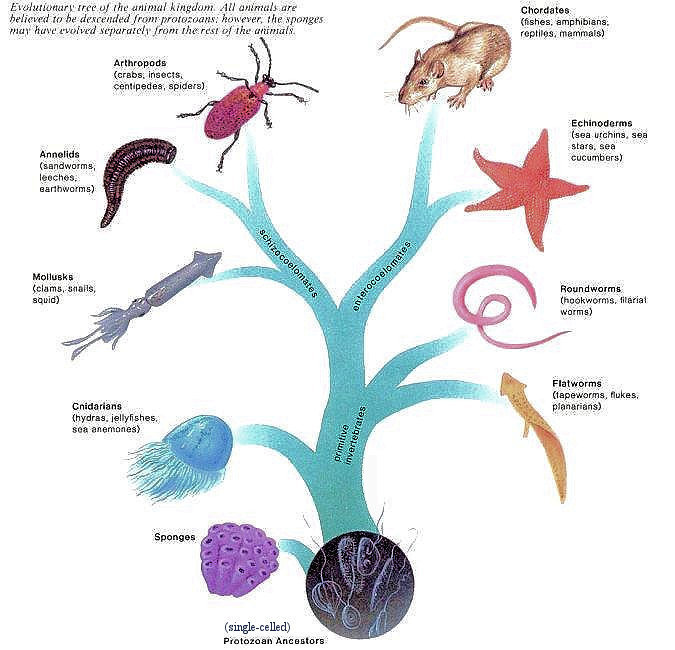
Acorn worms were traditionally classed in a group called the hemichordates, which along with the echinoderms (sea stars, basket stars, sea cucumbers, etc.) are  the vertebrates’ closest living relatives. As Colin Tudge notes in “The Variety of Life”, this is quite a remarkable thing.

In short, the chordates, which — in the vertebrates — include the most powerful, most swift, most versatile, and most intelligent creatures that have ever lived, emerged from amongst the ranks of animals that in large part are either sessile or seem to have a hankering to be so and some of which (notably the echinoderms) are without brains, not simply in the loosely perjorative sense but quite literally. Chordates, in short, have strange bedfellows. Nature it seems, likes to startle.

Together, these three groups — the acorn worms, echinoderms, and vertebrates (and possibly one or two other straggler groups) — make up the deuterostomes, a term you may have heard before. The characteristics that reliably define the group are not flashy features found in adults, but minute and highly conserved features of embryonic development (which are thus protected somewhat form the fiercest forces of natural selection) like anal pore formation, embryonic cell cleavage patterns, and larval types, so we will save that story for another day.

As per usual, the biologists are still fighting over what the exact relationships are between these groups. This diagram is as good as any for exploring it.

Most books describe acorn worms as creeping around or living in muddy or sandy U-shaped burrows on the sea floor. They are always depicted as frumpy and vaguely suggestive little creatures like the one pictured at top. When I taught introductory biology in grad school, our teaching specimens came from the same lot, and I remember dutifully fishing one of these not-so-impressive little creatures out of a jar to show my students.

And so the ugly larva grew up to be a beeeeee-uuutiful enteropneust. Photo courtesy of David Shale.

So — and here’s the point of this post — imagine my surprise when last week I opened up a slide show on wired.com of an expedition to either side of the Mid-Atlantic Ridge (run, do not walk,  to see) of new finds by British researchers in the Census of Marine Life aboard the RSS James Cook that was practically a debutante ball of brightly colored deep sea acorn worms. Worms with the frill and fuss of bustled, petticoated ladies. Worms that were observed swimming. Acorn worms, that, in short, look nothing like acorn worms. Homely burrowers my arse! Look at the diversity of color and form! On seeing these pictures, it is less hard to imagine the common ancestors of these organisms and us evolving into fish. But as always, these pictures beg the question: What else is awaiting discovery out there . . . ?

{ 3 comments }