The Hidden Fungi

by Jennifer Frazer on May 23, 2011

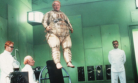
Rozella sp., a previously discovered internally parasitic cryptomycete totally devoid of a chitin cell wall. I think they're the brown truffley balls. Click to enlarge. Creative Commons Timothy Y. James. License at http://tolweb.org/Rozella_spp./103935


This post was chosen as an Editor's Selection for ResearchBlogging.org Unless you are a biologist, DNA can seem like pretty boring stuff: just an endless string of A’s, T’s, G’s, and C’s that functions as life’s hard drive. But to appreciate DNA if you are not a biology buff, you have to think big picture. You have to think of it as a tool — as a biological story detector. And last week, scientists announced that, with the help of DNA, they had stumbled on an amazing story. The fungi, it turns out, had a big secret.

To listen to news reports, you might have thought the big news was that the fungi have an interesting new set of oddball relatives. But these news reports completely missed what I consider the most earth-shattering news in the paper in Nature announcing the discovery, cleverly hidden at the end of this sentence:

Our analyses also recovered a highly diverse clade of environmental sequences branching with the fungi and demonstrated that current models of fungal evolution and biodiversity, which are largely based on cultured microbes, have missed a huge fraction of the kingdom (perhaps even approaching half).

Wait– hold up. HALF?!! Thus, the news isn’t that there’s some new oddball fungal relatives out there. It’s that the new oddball fungal relatives may make up half of Kingdom Fungi. And the first sample of this huge, undiscovered and massively diverse group came from none other than one of the scientists’ very own university pond. In spite of our best efforts to survey life, we had virtually no idea these things were out there — in virtually every environment we have now looked.

To understand how we may have been ignorant of half of Kingdom Fungi, you must think back to the time before DNA. To discover new life that couldn’t be seen with a hand lens, you had two options:

  1. Take some dirt or water. Wash and dump said sample onto a microscope slide. Apply various dyes as desired. Squash under coverslip. Place under microscope and stare intensely, searching for something that might be alive. Repeat thousands of times. See doctor about eyestrain.
  2. Take some dirt or water. Dump said sample into a petri dish containing some food-like substance. Wait. Pray the smell is not too bad. Place sample of stuff that grows on microscope slide. Squash under coverslip. Place under microscope and stare at similar-looking blobs. Repeat thousands of times. See doctor about eyestrain.

As you can see, this system left a lot to be desired. And it’s how hundreds of groups of organisms got mis-classified with other groups, because all biologists had to go on was the way the microbes looked and — in some cases, behaved — under a microscope. In addition, many groups of life on Earth are not culturable in a petri dish, or have evolved to look and behave in very similar ways, regardless of their ancestry.

Enter DNA. With this molecule, we can detect true heredity by comparing how the unique sequence of four letters in the code have gradually changed over time in related organisms. With millions of positions for letters in a defined order (like a cryptokey or license plate), it is easy to tell who is related to who and what is unique. By doing massive sampling of all the DNA in the environment, we can now easily sample for and pick out new variations of known genes (and the new organisms they must represent) in dirt and water samples, because they will look unique even if the organisms that make them do not*. And we can compare those new sequences** to sequences of organisms whose appearances and taxonomies we understand — in this case, a broad selection of fungi and their closest relatives.

AND we can use sequences unique to new groups as organismal metal detectors (scientists call them “probes”) to finger them under the microscope so we can see what they actually look like. Since DNA is a pair of complementary strands that bind to each other, you can make a probe that’s complementary to the particular sequence you’re looking for*** and attach that bit of DNA to a fluorescent molecule. Then you stick it in your hopelessly confusing sample of water or dirt and turn out the lights. Voila! Your organism lights up like a Christmas tree! No more eyestrain.

And that is just what a group of enterprising mycologists did. Biologists have sampled many different environments for DNA from all sources. In this study, scientists accessed online records of those sequences and looked for any that seemed to fit in with the fungi. They were in for a shock. As it turned out, there was an enormous group of organisms that branched with the fungi, that apparently no one had ever seen or cultured before, with the exception of one genus. Here is what they found:

This figure is actually much simpler than it looks. All the branches at the top with the colored dots represent organisms new to science in the new group, dubbed the “Cryptomycota”, or “hidden fungi”. There is one exception: a group of strange parasites called Rozella, which was already known. Everything else with the exception of the “opisthokont outgroup” — the group most closely related to Cryptomycetes and Fungi — are organisms from the known Kingdom Fungi. The rainbow of colors give you a feel for all the environments from which they found this new group. The only place they did not find cryptomycetes was seawater, although they did find them in mud on the seabed. The branch lengths should give you a feel for the diversity. It is incredible. The breadth of ribosomal DNA molecular diversity shown in they cryptomycetes here is similar to that of the known fungal kingdom.

What did these critters look like once fluorescently tagged by DNA? So far, the cells found in samples were 3-5 micrometers long — not much bigger than bacteria — and capable of making a microtubule-based flagellum like other good eukaryotes. In two of the three groups of cyrptomycetes they studied with probes, they found more than half of the cells had single flagella. Other, non-flagellate examples seemed to like latching onto — of all things — diatoms, the algae that make glass pillbox houses in a blinding array of uber-cute shapes. In addition, though they clearly group with the fungi, the Cryptomyctes — including Rozella — lack a very important marker of fungus-hood: they have no chitin/cellulose-rich cell wall.

For fungi as we know it, this is about as elemental as it gets. Their distinctive tubular, chitinous cell walls have enabled them to infiltrate a variety of foodstuffs that other creatures cannot and digest them osmotically. That is, grow into food that is concentrated in sugars or other nutrients that would make other cells shrivel up and die thanks to osmosis if they attempted it. We humans exploit this effect to preserve our food. Pickles, preserves, and peanut butter are so rich in salt, sugar, and general osmotic potential respectively that no bacteria or fungi can survive. But in nature, there’s a gray area where only fungi are bold enough to tread. They grow into their food (i.e. log, leaf, leftovers, etc.) They excrete digestive enzymes. They absorb the resulting goo. Life is good.

But the cryptomycetes have no such chitinous cell wall. The researchers know this because they stained the cells that probes identified as cryptomycetes with chemicals that bind to chitin and cellulose, the stuff of fungal cell walls, and they lit up like a Christmas tree during a power outage. Which is to say, they did not.

This is a problem for our definition of Fungi. As I learned it, to be a fungus requires five things:

  • you are eukaryotic (you have a nucleus where you keep your DNA and various other cute organelles orbiting it)
  • you are heterotrophic (you get food by taking it, rather than making it)
  • you are absorptive (we already covered this)
  • you reproduce with spores (a much better method than that used by mammals, IMO)
  • you possess a tubular body with a chitin cell wall

Yeast are somewhat exceptions to these last two; they are spherical and reproduce by budding. But they do not make up the majority of the fungi, by far, and so far, no one has ever bucked the chitin rule.

Hmmmm.

This brings us to the real questions wrought by this study: Are the Cryptomycota truly fungi? Or are they a larger group to which the fungi belong? Or are they a sister group of fungi? Did they ever have chitin cell walls or have they lost them? Are there vestiges of chitin in ephemeral life stages the researchers may have missed? There has already been some commentary on these questions in the comments here. We may learn a lot about the evolution of fungi based on the answers. One thing seems clear: if we decide they are “Fungi”, our definition — and our perceptions of the group — must change.

Aside from these important questions, we also have the delicious prospect of discovering how the contents of this new clade — the Cryptomycota — look and behave. There is almost no telling what we might find among some of the more eccentric members, though based on their fluorescence experiments so far, the paper’s authors suggest a saprophytic or parasitic lifestyle for the majority of species based on what they’ve actually seen under the microscope: a free swimming zoospore stage with a single tail, a resting cyst stage, and period of attachment and feasting upon a “second-party cell”, a.k.a. a helpless diatomic host or rotting microbial corpse (like leeches — or hyenas). But the group is so genetically diverse there are probably many, many ways of life. Based on what we know of the fungi, the authors suggest, there may be many life stages and accessories yet to discover: germ tubes, root-like food-harvesting devices called rhizoids, and spore-making houses called sporangia, filter-feeding structures, DirectTV dishes, etc.

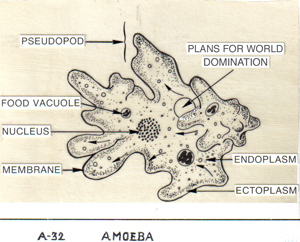
Without a cell wall, they may even be phagocytic — that is, able to engulf, swallow, and digest prey internally. Outside the Cryptomycota, fungi’s closest sister group is the nucleariids, a group of amoebae with filamentous (hmmm . . . ) pseudopods that feed, as amoebae do, by phagocytosis. Makes a girl think.

As the biologists say, this is an exciting time to study life. When reports like this come along, I feel a bit like a mapmaker in 16th Century Portugal hungrily studying the blank spots on the map. Can’t. Wait.

_________________________________________________________________

* because those groups will have two very different evolutionary histories, and thus, genetic patterns.

** in this case, of ribosomal RNA genes, a commonly sampled subject because they change so slowly and are present in everything

*** in this study, they used ten 18-base pair long clade-specific probes for taxonomic analysis fluorescent tagging purposes
ResearchBlogging.org
Jones MD, Forn I, Gadelha C, Egan MJ, Bass D, Massana R, & Richards TA (2011). Discovery of novel intermediate forms redefines the fungal tree of life. Nature PMID: 21562490

{ 7 comments }

Upcoming Colorado Fungal Learning Opportunities

by Jennifer Frazer on May 18, 2011

Can *you* ooze blood from your skin without the help of a friendly hemorrhagic fever virus? Didn't think so. This red fluid isn't actually blood, but I have no idea what it is. Hydnellum peckii -- "Strawberries and Cream" -- in the mountains of central Colorado near Winter Park. Author's photo.

So . . . you like what you’ve heard about fungi and want to learn more. And you live in the general Frontal Range area. Then have I got some opportunities for you: next Monday I will be giving the same lecture I gave in March at the Colorado Mycological Society (“The Many Ways to be a Fungus (in Colorado)”) for the Pikes Peak Mycological Society in Colorado Springs. See here if you’d like to come — the meeting is free.

Then, this August, I’ll be once again teaching “Wild Mushrooms” for the Boulder County Nature Association. It’s an evening lecture in Boulder followed by two field trips on Saturday mornings two weeks apart. Lots of fun will be had by all. $80, or $70 if you’re a BCNA member — find out more here.


{ 2 comments }

Falling in Love with Biology

by Jennifer Frazer on May 17, 2011

Most readers of this blog are probably biophiles of one sort or another, but it takes a special sort of passion (or lunacy) to start a blog on a subject in your spare time and write about it for hours on end without any expectation of renumeration. Yet I have done it now for over two years.

Why? A feeling, and the burning desire to share it with others. At its heart, my love of life on Earth is a love of shape, form, color, texture, and mechanism (which is why I put such an emphasis on good, large photos, diagrams, and art at this blog) — of the sort that’s put into overdrive by art like Haeckel’s drawings of ascomycete fungi at left, and of course, by seeing and understanding the organisms themselves. There’s also a joy that comes from looking at a pine tree and understanding (and being able to visualize) how that sucker works from root to shoot that I imagine is not far off from the feeling aerospace engineers have while drinking in a Saturn V rocket.

In fact, I recently read a description by a meteorologist of his similar feelings that convinced me I am not alone. The following is from the autobiography of meteorologist Richard Reed, who had an aptitude for science and math when he enlisted in the Navy at the outbreak of World War II but no idea what to do with with his life. He was previously planning to become an accountant when he signed up for the Navy forecasting service:

Freshly minted young ensigns Max Edelstein and Alvin Morris, the latter to become a longtime friend after the war, were assigned the job of teaching the trainees the elements of meteorology. To aid their instruction they suggested that we read a popular— and deservedly so—elementary textbook by Blair. I have never forgotten this experience. Once started on the book I could not put it down, staying up that night until I had finished reading it and feeling at the conclusion that I had thoroughly absorbed the material despite my relatively weak scientific background. If there ever was a case of love at first sight for a scientific subject, I experienced it that day (and night). There are those who view unusual ability in math and physics as the key to scientific success and its manifestation in a particular subject as largely a matter of accident. I have never subscribed to this view. The aesthetic feelings aroused in me by weather patterns and the fascination I felt for weather phenomena as physically evolving entities have always seemed to me inborn facets of my being. I cannot picture any other field of study having had the same emotional effect.

If you find something like this in your life, grab it and don’t let go. I once saw a documentary about a jazz musician (I cannot remember which one) who went through the rise to stardom followed by de rigeur drug- and alcohol-induced crash. At the peak of his fame, he had played in an exclusive jazz club. Now that he was penniless, he sat outside it on the curb, playing the same songs he had done before for free. He could not imagine anything else he’d rather do with his time, whether he was being paid or not.

When I watched this, I was in college and struggling with what to do with my life. For four years after I realized I had no desire to be a research scientist after all, I had no clue. If only, I thought, I could find a career I felt about the way that jazz guy did. Something I was so passionate about that I would do it for free. And eleven years later, here I am, blissfully, unemployed-ly, doing what I love and giving it away. I am thinking perhaps that life has now provided me the cosmic kick in the pants to go for it fully — and maybe even get paid.

So in the wake of my recent job loss, I will be embarking on the grand adventure of the freelance lifestyle. I am excited about this change; it is one I’ve been hoping to make for a long time. At the same time, it’s also terrifying. The mortgage, health insurance premiums, and grocery bills must all get paid each month. And so I will continue to look for part-time work. But I have a deep-down feeling that this is the place, and now is the time. In addition to this blog and to magazine, newspaper, and internet work, I am hoping, one day, to also write books that will also help you fall in love with (and laugh a bit about) some of your planet’s lesser-known co-passengers. And I am hoping you will want to read them. : )

Thanks to all who have sent in freelance contacts so far; more are most welcome. And thanks for everyone who’s advised me to seize the moment. Fortune favors the bold, so the Romans said, and so I hope.

{ 11 comments }

The 5-Million Year All-You-Can-Eat Buffet

by Jennifer Frazer on May 12, 2011

Algae having a par-tay today. It's nothing compared to the 5-million year bender they went on 250 million years ago. Creative Commons eutrophication&hypoxia

This post was chosen as an Editor's Selection for ResearchBlogging.orgAbout 250 million years ago in what is today the vast backwater of north central Siberia, the Earth coughed forth an unimaginable quantity of lava over the course of 1 million years. The liquid rock was a low-viscosity, thin stuff (for lava), so instead of forming a field of towering volcanoes it oozed out into endless plains. Covering some 1.5 million square kilometers today (600,000 square miles — something like the surface area of Europe), the beds may have originally covered 7 million square kilometers and taken up 1 to 4 million cubic kilometers in volume.

Scientists still do not agree why this happened, although it has happened many times before and since around the world. But they do agree on the timing: it happened at the same time as the Great Dying — during the Permo-Triassic extinction just prior to the age of dinosaurs that wiped out more life on Earth than any other, including the one with the giant asteroid with our name on it.

Not all scientists agree that the Siberan Traps, as they are called (trap comes from the Swedish word for “stairs”, which is how the frozen lavas can appear today) chiefly caused the extinctions. But many do, and it seems to be solidifying (er, pardon the pun) as the majority opinion.

What is also known is that life took some time to recover from this cataclysm. For at least 5 million years after, we can find little in the fossil record. Scientists have wondered whether this was a case of too much or too little food.

How, you might ask, could too much be a problem? Well, visit your nearest pond contaminated by fertilizers from lawns, golf courses, or farms and you will see: vast swarms of algae, that hog all the oxygen and choke out “higher” forms of life like fish. Or, for that matter, the New-Jersey-sized Dead Zone in the Gulf of Mexico, which formed in response to the endless streams of fertilizer runoff from farms pumped into the sea by the Missouri, Mississippi, and Ohio River system. We call this eutrophication*, and now, the authors of a paper in in Earth and Planetary Science Letters are pretty sure it is what prevented most life from recovering for those five million long years.

What happens when the party gets a little too wild. A lake in China with rampant eutrophication. Creative Commons eutrophication&hypoxia

How might scientists figure this out? As it turns out, one of the enzymes that catalyzes photosynthesis has a quirk. Thinking back to high-school chemistry, recall that the nucleus of an atom is made of protons and neutrons. Different elements are defined by the number of protons their nuclei contain. Carbon *always* has six protons. If it had seven, it would be nitrogen. But elements can vary in the number of neutrons they have. Carbon, for instance, can commonly have six neutrons or seven. Carbon-12 represents the former, while carbon-13 the latter.

As it happens, one of the chief enzymes of photosynthesis — RuBisCO, the one responsible for grabbing carbon dioxide from air and setting it on the path to become glucose — processes carbon dioxide containing carbon-12 a little faster than carbon dioxide containing carbon-13. Over jillions of cycles, the difference accumulates, and life becomes enriched in carbon-12. It makes little or no difference to the organisms themselves.

But it makes a big difference to scientists, who can use this knowledge to tell how fast the oceanic biological pump — the transfer of carbon from the atmosphere and surface waters to the seabed by microorganisms that live, die, and sink — is churning. The more these microbes nom, the more carbon-12 builds up in preference to carbon-13 in seabed deposits, where marine algae sink, store carbon, and are eventually pressed into limestone after they die. Scientists who studied marine deposits recorded in Chinese rocks during this 5-million year gap have found the carbon-12 enrichment is about double what exists today. If life had starved after the Traps did their dirty work, they would expect to see the opposite.

This is the scenario the authors of this new paper believe may have happened: as they erupted, the Siberian Trap lavas and the rocks they metamorphosed by contact spewed carbon dioxide (as well as many other volcanic gases) into the atmosphere. This warmed the atmosphere, which increased the evaporation of water, itself a greenhouse gas, perpetuating the cycle.

At the same time, more water vapor produced more rain, which weathered the land quickly. All that runoff drove incredible quantities of phosphate — a nutrient that limits the rate at which marine algae can grow — into the ocean. With a new all-you-can-eat buffet of phosphate and carbon dioxide at their disposal, marine microbes went nuts. The ocean was stripped of free oxygen, preventing any animal life that had managed to survive the extinction itself from regaining ground. Once the initial trauma of the Permo-Triassic extinction was over, these algae must have bloomed in quantities unimaginable. What is now the Blue Planet, and once may have been the White Planet, was briefly, it seems, the Green Planet.

What finally stopped the madness (from our perspective as vertebrates), was the waning of the volcanoes. Eventually, carbon dioxide levels dropped, and warming and rainfall decreased. With less erosion, oceanic phosphate and nitrogen concentrations dropped. Without enough of these nutrients to go around, algal numbers shrank, leaving enough oxygen around for other forms of life to exploit. And, luckily for us, they did.
___________________________________________________

* Which is the reason you should all be using phosphate-free detergents.
ResearchBlogging.org
Meyer, K., Yu, M., Jost, A., Kelley, B., & Payne, J. (2011). δ13C evidence that high primary productivity delayed recovery from end-Permian mass extinction Earth and Planetary Science Letters, 302 (3-4), 378-384 DOI: 10.1016/j.epsl.2010.12.033

{ 2 comments }

Hot Rhodopsin

by Jennifer Frazer on May 5, 2011

I would like a fascinator made in this shape. The dinoflagellate Oxyrrhis marina with its transverse and longitudinal flagella. Creative Commons Census of Marine Life E&O

Life on Earth is full of weird convergences. For example, the cell walls of fungi are made of chitin, as are the shells of insects and arthropods like lobsters and crabs. Why? Whether due to chance or something deeper, I have never heard a good explanation.

Similarly, the proteins called rhodopsins are found in two seemingly completely unrelated places: the retina of your (and all vertebrate) eyes, and in some photosynthetic archaea and bacteria. We use ours for detecting light (it’s in the rod cells used for low-light vision); they use theirs to pump protons (as illustrated and discussed recently here) to make food.

And now, scientists have discovered a predatory dinoflagellate that has apparently stolen a bacterial photorhodopsin from one of its meals — and is using it (see also here).

The protein in question is called rhodopsin (Greek root “rhodo” = rose and “opsis” = sight) because it absorbs blue-green light and so appears purplish-red. Rhodopsins have seven protein coils called alpha helices that pass through membranes the protein is embedded in. In turn, embedded inside the helices is retinal, a light-sensitive pigment. As a result, the whole structure changes shape in response to light. In vertebrates, rhodopsin has another subunit called a “G protein“. G protein acts as signal transducer, or on-off switch in a signaling cascade — just like a switch in an electrical circuit. Vertebrates use it to relay information to the brain about what the eyes are seeing. Bacteria use their rhodopsin simply for pumping protons, not for signalling, so they have no need for G protein section.

The bacterial protein — still seven alpha helices and a retinal — resembles overall shape of vertebrate protein, so scientists suspect they may be distantly related. Proteorhodopsin — the bacterial version, was only discovered in 2000, but the (now ironically named) bacteriorhodpsin found in archaea was discovered much earlier, in archaea living and photosynthesising in sun-drenched salt flats, brackish pools, or salt marshes.

The dinoflagellate Oxyrrhis marina (ox-EE-ris MARE-i-na, I think) is neither a vertebrate nor a prokaryote (bacterium/archaeon). And yet O. marina makes so much rhodopsin it has turned pink. How? Scientists believe the gene for the proteorhodopsin was acquired by what biologists call horizontal gene transfer — that is, O. marina was happily digesting its photosynthetic bacterial prey when the gene for this protein somehow wandered off and found its way into the nucleus (where DNA is stored) and slipped, spliced, or got sliced into the chromosome.

This appears to be a case of “hedging your bets” because O. marina is now both photosynthetic and predatory. So in this case, you can make your own cake and stalk it too. Scientists believe they use this protein not only to make energy by pumping protons and passing them back down a chemical gradient via an ATP synthase, but also to help digest the very prey they stole it from. As they say, all too easy.

But before I say more, a few words on what dinoflagellates are are in order.

The dinoflagellate Ceratium longipes, in all it's tricorne glory. One, and possibly two flagella visible. Creative Commons Census of Marine Life E&O

In short, dinoflagellates are two-tailed plankton. They are also protists, the loose association of single-celled organisms with DNA inside nuclei and cellular organelles that are usually much bigger than bacteria or archaea. About half are predatory, half make their own food, and obviously, now we know some do both. The photosynthetic lot are the second most abundant constituent of the photosynthetic marine plankton after diatoms (which I covered here).

Dinoflagellates (from the roots for “whirling whip”) are also alveolates like ciliates (including the paramecia I wrote about here) and apicomplexans (which include Plasmodium, the protist that causes malaria). That means they often have sacs called alveoli under their cell membranes, trichocysts (defensive spikes that shoot out like harpoons), and tubular mitochondrial folds, or cristae. Some even produce structures like the nematocysts of jellyfish. You can explore how dinoflagellates fit into the life family tree here (use the black arrows to move toward the root).

Some dinoflagellates cause the poisonous red tides infamous for sickening fish and swimmers alike. Others build up in tropical fish or shellfish and cause ciguatoxin or paralytic shellfish poisoning in people. Others become the zooxanthellae, the green algal symbionts found in coral and other animals and protists. These photosynthetic dinoflagellates vastly increase the speed at which corals can build their skeletons. And finally, some dinoflagellates (including some red tiders) are bioluminescent, emitting short flashes of light when disturbed, and are responsible for the unearthly glow of the wakes of ships passing through tropical waters in the night.

Bioluminescent red tide dinoflagellates getting all bright and bothered by rolling surf. Creative Commons Catalano82. Click for link.

Dinoflagellates have some interesting quirks; their chromosomes stay condensed throughout the cell cycle and down relax into a spaghetti pile of chromatin (individual DNA threads) between cell divisions. And they don’t wrap their DNA around proteins called histones as most of the rest of us upstanding eukaryotes do. They prefer instead to attach it to the inside of the nuclear membrane. Also, they tend to eschew the reliable DNA base thymine in favor of the boutique 5-hydroxymethyluracil.

My plans to take over my neighborhood swimming pool are now complete . . . Dinoflagellate blueprints. Creative Coimmons Shazz. Click image for link.

Their two flagella — which emerge from the same point — are often set in grooves. One is a belt-like transverse groove called the cingulum, and the other is a longitudinal groove called the sulcus. You can see those grooves in a beautiful scanning electron micrograph here. (See also here and here for a dinoflagellate that I swear resembles Baron Harkonnen in his fat suspensor suit). The flagellum set in the cingulum wraps all the way around, while that set in the sulcus trails off the back like a rudder. Strangely, the transverse flagellum is responsible for most of the forward motion and is what sets dinoflagellates whirling, while the longitudinal chiefly helps steer.

Dinoflagellates are also really good at engulfing photosynthetic organisms and endosymbi-izing them. Most of their chloroplasts have three membranes, suggesting they came from an ingested alga, not a bacterium, and are the result of a secondary engulfment, or endosymbiosis. Others have chloroplasts of different color, shape and form, some still with nuclei. Thus we can infer that dinoflagellates are really good at finding themselves new photosynthetic dates by hook or crook — definitely crook, in the case of Oxyrrhis.

Flexibility and Finding a way seem to be themes with dinoflagellates. O. marina, for example, is common in shallow tide pools around world; it will go so far as to cannibalize its own species if it comes to it and can take down prey almost as big as it is. Photosynthesis. . . hunting . . . cannibalism — this thing is the MacGyver of protists. It’s going to survive with whatever comes to hand. So if you’re ever doing O. marina research, please do us all a favor: do not keep bubble gum, paper clips, or high explosives anywhere near your experimental organisms. Thank you.

{ 2 comments }

Seeking Science Writing Gigs

by Jennifer Frazer on May 2, 2011

Connections -- here's hoping I've got some. Fungal mycelia from a bathroom remodel. Creative Commons Bob Blaylock. Click image for license.

Dear Readers,

I have a bit of sad personal news. I learned today that my day job has been cut due to a budget shortfall. As a result, I am back on the job market. I have six years of experience as a health and environment reporter and science writer, and in 2007 I won a AAAS Science Journalism Award. If you like my work and know of any science writing job possibilities or freelance work that I could do from Colorado, please get in touch with me via the email address on my Portfolio page. Alternatively, you can post a comment if you don’t mind a few hundred other people seeing it. : )

Regardless of these circumstances, I plan to keep this blog up and running. Fortuitously, my post “Bombadier Beetles, Bee Purple, and the Sirens of the Night” was selected by Ed Yong as one of his “Science Writing I Would Pay to Read” posts for this April. If you’d like to make a donation to all the science writers (including me) who were featured this month there, you can do so via a paypal button that says “Support Science Writers” at his blog.

Thanks in advance for any help, advice, or moral support. : )

Jennifer

{ 8 comments }

Sightseeing the Deep Sea

by Jennifer Frazer on April 28, 2011

In case you missed it earlier this month, Sir Richard Branson is building a new one-person sub to explore five of the deepest points on Earth. When you are certifiably the coolest man on Earth, you can do stuff like this . . .

He has thus partially granted my wish from last year, and even plans to use the missions for science as well as exploration and adventure. . . but Richard, we’re only half-way there! Once you’re done planting Virgin Oceanic flags all over the seabed, please build a commercial version of this baby to take the rest of us down there! I’m willing to pay as much as the projected lowball cost to the space cadets going up on Space Ship Two — and this ride doesn’t even require escaping any pesky gravity wells via loads of expensive, atmosphere-heating rocket fuel. Have I sold you yet? Fingers crossed.

Here’s a similar plea from Dr. M over at Deep Sea News, although it looks like, sadly for him, the current model is a one-seater.

{ 1 comment }

The Weird Wonderfulness of Life Inside You

by Jennifer Frazer on April 26, 2011

Though we don’t think about it very often, there’s a universe of amazing life humming along inside you. For example, inside nearly every living non-bacterial cell in your body, you will find …

This remarkable video is one of a series recently produced by a biological animation initiative at Harvard. Earlier, they made a more extensive movie of the inner workings of a cell (See the adventures of a white blood cell in  The Inner Life of the Cell here, and the considerably less inspirational but more enlightening narrative explanation of the video here (this is what happens when science writers do not write the script)). They’re not new; Carl Zimmer has posted one of these videos before, but I wanted to make sure none of you missed them. They’re a far cry from the cartoonish still drawings (see below) we had to use our imagination to envision in real life when I was in school.

So what’s actually going on in the mitochondrial video? Well, I’m not sure of every detail, but as a former biochemistry TA, I can give you a lot of educated guesses. The movie appears to begin with mitochondria — sub-cellcular power stations — inching along a part of the cellular skeleton called microtubules. The job of mitochondria is to finish the process of turning the energy stored in the bonds of glucose into useable power through respiration. As energy is harvested from the electrons prised off glucose, they are finally passed to oxgen, which accepts them along with some hydrogen ions to form water. Meanwhile, the carbon that was tied up in glucose ends up as carbon dioxide. This forms the basis of your inhalation of oxygen and exhalation of carbon dioxide with each breath you take. Anyway, back to our film.

Then an (?)amino acid chain (fragment of a protein) of some sort escorted by (?)chaperone proteins enters a mitochondrion via a pore through its double membranes*. Then you see the contents of the mitochondrion: an asteroid field of colorful proteins zipping about amid strands of the mitochondrion’s DNA.

Then, looming in the distance like the Pillars of Hercules (or the warship encrusted columns inside the motherships in Independence Day — which seems to be the point. They are going for cinematic here.) are tubular mitochondrial cristae, or folds of the inner membrane. These folds increase the surface area available for respiration.

Embedded in these columns are the rotary engines of mitochondria — enzymes called ATP synthases. These proteins are fascinating feats of natural selection that rotate as they charge their substrates (the molecules they will act upon): namely, ADP. Swarming around these proteins like fireflies are hydrogen ions (H+ — essentially, a proton, but often surrounded by three oxygens in aqueous solutions) generated by stripping glucose of electrons like a car in chop shop.

Without going too deep into the gory details, when a cell burns glucose, it performs some preliminary reactions in the cytoplasm (glycolysis) and sends the remaining energy-bearing bits into the mitochondrion for full processing. After enzymes performing the Citric Acid cycle in the interior of the mitochondrion (called the matrix) squeeze more power out and release what’s left of the glucose as carbon dioxide (CO2), the electrons glucose has yielded are passed down the electron transport chain of proteins embedded in the inner membrane, which use the energy thus released to pump hydrogen ions out of the matrix into the space between the mitochondrion’s two membranes. I think you can see this happening at about 1:08.

The inner membrane, unlike the outer, is highly impermeable to most molecules — even to tiny hydrogen ions. The resulting ionic gradient can only flow back downhill through a rotating pore in ATP synthases**.  The passing hydrogen ions powers their rotation and their charging of ADP to ATP, the cell’s energy currency (an explanation of this fascinating mechanical process can be found here under “binding change mechanism”). It’s such a clever system that engineers have designed engines (called the Wankel Engine) based on the same principle and built them into working cars — namely, the Mazda RX-7 and RX-8. I know this because one of my biochemistry professors at Cornell had actually owned one of these for that very reason (You know you are a nerd when . . . ).You can see the biological version of this process happening at about 1:15, where small molecules enter the head of the synthase, light up to let you know they’ve been charged, and then are released to please go play nicely with the rest of the cell.

Here is a conventional representation of what I just described — I, II, III, and IV are proteins of the electron transport chain, NADH is an electron ferry that shuttles said particles from ex-glucose pieces to the electron transport chain, and succinate is an intermediary in the citric acid cycle (for those that remember, this is the step that generates FADH2):

During all this action in the movie, the camera also passes once or twice through the undulating lipid bilayer of membranes, where the kinky double-tailed (and faintely spermish) phospholipids jostle against each other to keep the membrane fluid. Mitochondrial membranes actually contain many fewer sterols (cholesterol is one — they are molecules that help stabilize membranes) than the cell membrane, giving the mitochondrion greater shape-shifting powers.

I think the next-to-last scene is an ATP/ADP transport protein that actively shuttles ADP into the matrix and ATP out. Finally, you see all the mitochondria swarming toward some big shiny thing (centrosome? endoplasmic reticulum? Who knows! Scene list please, Harvard!) like star cruisers converging on a galactic rendezvous. Actually, mitochondria do sometimes cluster near where they are most needed. For example, in cells with flagella, they may cluster near the base of the tail.

All in all, the mitochondrion’s a tightly run ship. Lest the ID community use these incredible little machines as evidence of “stasis” or “irreducible complexity”, let it be known that anaerobic (non-oxygen breathing and non-mitochondrial) bacteria alive today have proteins almost identical to ATP synthase that function in reverse: powered by ATP, they serve to detox the bacteria of H+ to rid them of the acidic by-products of the less-efficient but still-better-than-nothing energy-producing process of fermentation. The cytochrome complexes (aka I, II, III, and IV, the cogs of the electron transport chain) may have evolved for similar detox purposes in other ancient bacteria before being combined with ancient ATP synthase by natural selection to form the well-greased respiratory engines we have today.

Typical plant and animal cells contain hundreds or thousands of such mitochondria, though their number ranges from one bizarro giant in a few single-celled protists to several hundred thousand in well-provisioned egg cells. I’m not certain why the directors of this film chose to show this mitochondrion with tubular cristae. Most vertebrates have regular laminar, or sheetlike, cristae (remember that it was unusual that alvaeolates (the paramecia, ciliates, dinoflagellates, and apicomplexans) had tubular cristae), though plants have both sheets and tubes in their mitochondria, and are more irregularly shaped and sized.

Of course, proteins inside mitochondria don’t really float around looking like someone blew up a box of Trix in the space station. In reality, my understanding is they’re all sort of, well, clearish at that scale. And in this thorough NYT article on recent advances in molecular animation, scientists acknowledge that molecular animators also take liberties with space.

“Some animations are clearly more Hollywood than useful display,” says Peter Walter, a Howard Hughes Medical Institute investigator at the University of California, San Francisco. “It can become hard to distinguish between what is data and what is fantasy.”

But clearish molecules and vast distances would make for a pretty dull movie, so I don’t begrudge them their colors. This situation reminds me of the Peter Jackson Conundrum: was the Lord of the Rings better before your head was filled with Peter Jackson’s version of everything? And wasn’t it better when only the people who actually took the trouble to do all the reading were in on the magic?

My gut feeling is that these movies are a good thing, as is sharing the wonder with the masses. If we wish to make the case to society that science is important and worthy of time and money even on its own terms, animations like these help. It is also unquestionably cool to see it all in such detail — revealing things we could not easily foresee without seeing everything together in glorious living color — even if our imaginations are a bit impoverished for it. It’s a worthy sacrifice, in my opinion, if our appetites are whetted.

________________________________________________

* The outer membrane is a product of a long-ago engulfment of a bacteria by a predatory cell — the inner membrane is the ancient bacterial membrane and the outer membrane is the erstwhile vacuole. Further evidence for endosymbiosis includes that mitochondria (and chloroplasts) have their own single, circular (like most bacteria) DNA-based chromosome without a nuclear membrane from which they manufacture their own proteins and bacterial-sized ribosomes (which can even be interchanged with bacterial ribosomes in some cases) and replicate by division. They’re bacterially-sized too: 1.5 by 2-8 micrometers.

** In high-magnification photos of mitochondria, you can actually see the ATP synthases poking into the matrix like lollipops.

{ 1 comment }

The femme fatale Photuris. Photo by Bruce Marlin; click image for Creative Commons license and image source.

If I read my notes correctly, Thomas Eisner once had a pet thrush named Sybil who rejected only five insects out of the hundreds the entomologist offered her. They were all beetles. And one of them was a firefly.

For any other bird owner, this observation would have simply limited their pet’s meal options. But this was Thomas Eisner — one of the great entomologists and chemical ecologists of the 20th century. To him, it was a tantalizing clue, and he decided to find out what made the fireflies have all the thrush plate-appeal of haggis. What he stumbled onto was one of the great new natural history stories of the 20th century — and the latest in a string of Eisner’s greatest hits.

I know this because in fall of 1998 I was a student in BioNB 221 — Introduction to Animal Behavior — at Cornell University. Eisner, a professor at Cornell, taught the last six or so lectures, which I still have preserved in my notes. What I did not know at the time, and did not learn until afterward, was that Eisner was one of the great biologists of the 20th century. I found this out in later years, when his discoveries were featured in many an article at the New York Times, where I had a mysterious feeling of deja vu.

What I did know at the time was that I could not take my eyes off the screen while he was lecturing. I’m a fan of a good natural history story, which you may perhaps have gathered. Eisner — who was once E. O. Wilson‘s college roommate — was overflowing with them — and in many cases, because he’d figured them out himself. Sadly, Eisner died March 25. You can read more about his life in this fine remembrance by NYT reporter Natalie Angier*, whose daughter was lucky enough to inherit the contents of Eisner’s old burlap field bag and was, frighteningly to me, born around the time I sat listening to Eisner’s lectures. Angier wrote about his life. I want to share with you a few of the natural wonders I learned from him, sitting rapt in the darkened Uris Hall auditorium.

This Means War

Eisner’s specialty was the world of chemical warfare among plants and insects. Insects produce, steal, and reuse chemicals from plants and each other constantly. Millipedes can deploy hydrogen cyanide, whip scorpions acetic acid, and ants formic acid, but for Eisner, the poster child for entomological chemical defense was the bombardier beetle. “If you live on the ground,” he said, “you must either take flight quickly or defend yourself instantly.” The bombardier beetle went with option B.

The beetle takes chemicals called hydroquinones, mixes them with hydrogen peroxide and catalytic enzymes (peroxidase and catalase) in a reaction chamber in its hinder, and uses the resulting explosive formation of benzoquinones and heat to persuade frogs, ants, and spiders that their best meal options lie elsewhere.

Using grainy films he had shot himself, he showed us how beetles touched with probes could deploy a vicious defense with pinpoint accuracy in nearly any direction. He suspended the beetles over pH paper, so the 100°C benzoquinones they released would reveal their precision firepower.

This British film (which seems to have been created by intelligent design advocates who tried to abuse Eisner’s research for their ends**, so ignore the bit at the end. I couldn’t find another version, unfortunately.) incorporates some of the movies I saw that day, as well as explains how the beetle uses physical barriers to control its chemical defense system. I think you can even see Eisner in one of them for a few seconds at the end — he’s in the foreground.

And here’s David Attenborough describing the beetle in HD:

Don’t Feed on Me

Plants, too, load up on poison in hopes of warding off the hungry crowd. Nettle spines are filled with irritating chemicals, as are the latex canals or resin canals of flowering plants and conifers, respectively. Some plants store poisonous chemicals in their tissues like caffeine or nicotine, which in spite of their uplifting effects on humans, are actually insecticides.

But some insects have picked up on this gig, and begun using it to their advantage. Sawflies slice into the resin canals of pines and steal the sticky sap, storing it in special sacs for defense against ants. Monarch butterflies sequester milkweed toxins from their food, rendering themselves distasteful to predators. Assassin bugs coat their eggs with the noxious excretions from camphor weeds. Their young then reuse the chemicals for defense and to catch prey. We do this too, Eisner pointed out, by stealing the defense chemicals from fungi and other bacteria. We call them antibiotics.

Eisner told us of plant chemicals stolen and presented as nuptial gifts among moths, where female choose males whose flirting, aromatic antennae tell them they have stored the most alkaloid derivatives. That implies the male is both fittest and has the most to give to the pair’s offspring. For if the female mates, the male will transfer not only his sperm, but his alkaloid collection, which the female will carefully store with her eggs for the use of her young. Other moths do the same with salts they siphon from puddles.

And he told us of the evesdropping of kairomones — chemicals that, unlike pheromoes, used for intraspecies communication (like the moths), or allomones, which benefit the emitter of an interspecies pair (like the benzoquinones of the bombardiers or the stinking of skunks), benefit the receiver and betray the emitter. Think, for example, of the carbon dioxide that gives you away to mosquitoes; any scent, really, that betrays prey to predators can qualify. Eisner called it a “chemical gestalt”, the effect of “inevitable chemical leakage”. But the tables can also be turned. Predatory rotifers called Asplanchna unwittingly emit chemicals that alert prey rotifers called Brachionus to grow defensive spikes (read more about rotifers from this blog here and here).

One of my favorite Eisner stories, one that has especially stuck with me all these years, was about true bugs entomologists were attempting to rear in petri dishes on damp paper towels. The bugs’ development was, however, stalling; they could not be coaxed to adulthood. The scientists were baffled. Until, that is, someone noticed the paper towels were made from balsam fir, a tree that emits allomones to stunt insect development. This chemical was, apparently, surviving the paper-making process and continuing to thwart the trees’ insect enemies — even in death.

Bolas spiders use imitation pheromones — another allomone — to lure male moths in search of a date (the females, apparently, are immune to the spider’s charms). This video depicts the unfortunate result:

You may have heard of parasitoid wasps — the Alien-style predators of spiders, caterpillars and other insects that lay their eggs in their prey, where the young maggots proceed to devour their hosts’ organs while still alive before finally using their hosts’ spent husks as pupae from which young wasps emerge. But perhaps you did not know that some plants injured by caterpillars or aphids call out chemically to parasitoids to defend them. But the story gets better; the immune system of the host in some cases is destroyed by viruses injected by the parasoitoid wasps along with their eggs. “And(I underlined this in my notes) the viruses have also been incorporated into the wasp genome.” To which I further wrote, “1 organism now? Whoa.”

He told us how mammals, too, use pheromones. Babies can distinguish their mother’s milk from others, he said, and the scent of male armpits can regularize erratic female ovulation. In mice, the scent of strange male urine blocks implantation of fertilized eggs in female mice; the effect and reason may be similar to an article I just saw last week about mares aborting fetuses to save themselves investing energy in foals likely to be killed by rival stallions anyway. This could explain the spectacularly high miscarriage rate in mares (around 30%) who are trucked out to mate with top stallions but housed while pregnant with other males. That this is likely to have not one whit of effect on the way breeders practice horse husbandry is testament to the often hidebound thinking of humans.

The Bee-Letters of Flowers

But on top of all this research into chemical crossfire, Eisner also dabbled in the world of light and visual communication. Those who have studied physics know the electromagnetic spectrum of which light is a part is a vast array of energy. Earth’s atmosphere filters much that arrives, and most of what makes it through falls in the 320 to 2300 nm range. What we perceive as visible light falls in the 380 to 750 nm range. But that leaves a large part of the spectrum invisible to us. What if other animals could see different colors or different parts of the electromagnetic spectrum? As it turns out, they do.

We cannot see ultraviolet. But, through experiments worked out by a whole host of Germans, we know bees do. Conversely, bees cannot see red. Their vision lies in the 340 – 650 nm range. Blue, red, and green are the human primary colors. But the bee primary colors are yellow, blue, and ultraviolet. That implies there are a spectrum of colors that they see that we cannot. My mind bent a bit as I heard this — there’s a whole world of color out there that we can’t see!

And those colors needed names. Yellow + blue we can see along with bees — we call that blue green. But what about blue + ultraviolet? That was dubbed “bee blue”. Yellow + ultraviolet? “bee purple”. And, as it turns out, flowers are adorned in these shades, invisible to us but brilliantly displayed for bees. Flowers probably first used UV-absorbing pigments as sunscreen, Eisner said, and only later turned to them to decorate their petals. Now, bee blue and bee purple form pollen guides for bees, often flecking the tips of flowers and leaving a yellow disc in the center as a bullseye. You can see the effect in this photo collage of black-eyed susans with ultraviolet tips and a yellow center, though the bee would see both yellow and ultraviolet simultaneously as bee purple.

Cucumber flowers in natural light(left), and in ultraviolet falsely colored yellow(right). To bees, the flowers would appear bee purple with a yellow target -- the pollen guide. Creative Commons kevincollins123. Click for license and link.

The inability of bees to see red means that pink are red flowers are almost never pollinated by bees. On the contrary, only butterflies and hummingbirds — which are not red-blind — are attracted to red flowers. Eisner wrote papers about his experiments in this world as well, examples of which you can see here and here.

Which brings us back to what is likely his most famous experiments in light communication — the Tale of Photinus and Photuris. Following up on the expectorated clues provided by Sybil, Eisner extracted chemicals from the fireflies with various solvents. He discovered that the firefly she spat out — Photinus — contained a steroid called lucibufagin. When fireflies are caught, they “bleed” hemolymph full of this chemical. Spiders who catch and taste them let them go. They even release fruit flies merely painted with the chemical, the scientists discovered. Eisner found Photinus was chock-full of the chemical right from the start of the season. A larger firefly, Photuris, also contained this chemical. But only the females. And only later in the season. He began to glimpse the truth of a dark story.

Male fireflies searching for females make a species-specific pattern of flashes. Females respond with a single blink, but with a species-specific time delay from the male call. Photuris, coveting the chemicals of Photinus, imitates that response. When the male lands thinking he is about to get lucky, he gets eaten instead, and the female accumulates the chemical that allows her to escape predation by spiders and yes, thrushes.

How could one man do and learn so much? Perhaps because he never let the Lab get in the way of Life. This passage from Angier’s piece, in particular, explains why I love Eisner — and to a large degree why being a modern biologist was not for me.

Ian Baldwin, a professor of molecular ecology at the Max Planck Institute for Chemical Ecology in Germany, who studied with Dr. Eisner  in the 1980s, said of his mentor: “He articulated the value of natural history discovery in a time of natural history myopia. We train biologists today who can’t identify more than four species, who only know how to do digital biology, but the world of analog biology is the world we live in. Tom was a visionary for nonmodel systems. He created narratives around everything he did.”

In today’s “shiny polished science world, he was proof that there is no experience that can substitute for being out in nature,” said Dr. Berenbaum. “It’s classy, not low-rent, to stay grounded in biological reality.”

Thank you for the stories, Dr. Eisner, wherever you are.

____________________________________________________________
*My answer to the question of “Who are your favorite science writers?” with “Natalie Angier” probably terminated my interview for a science journalism internship at a major daily newspaper about seven years ago. The editor seemed to lose interest in me at that point. I wasn’t going to lie, and I’m still offended on her behalf.

** Indeed, they have also done so to my graptolites post. They linked to my blog post as part of a post using graptolites as proof of “stasis”.

{ 14 comments }

The Many Ways to Be a Fungus (in Colorado)

by Jennifer Frazer on April 16, 2011

At last, here’s a recording of the 45-minute lecture I gave in March to the Colorado Mycological Society: The Many Ways to Be a Fungus (in Colorado). The lecture discusses fungal diversity using Colorado examples, but it should be interesting to anyone who wants to learn more about fungi. I hope to have a full post ready for you early next week. In the meantime, enjoy!

The Many Ways to Be a Fungus (in Colorado) from Jennifer Frazer on Vimeo.

{ 0 comments }